Page 27 - ebook
P. 27

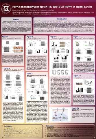
HIPK2 phosphorylates Notch1-IC T2512 via FBW7 in breast cancer


                                                                   School of Biological Sciences and Technology, Chonnam National University, Yongbong-dong, Buk-ku, Gwangju, 500-757, Republic of Korea


                                                                   *Corresponding author : Hee-Sae Park, e-mail : proteome@jnu.ac.kr


                                                                     Abstract                                                                                                                                                                                                                                     Introduction


                                                                                                                                                                                    Notch1 is a highly conserved transmembrane protein that plays a crucial role in cancer development, cancer cell survival, prolif- eration, and differentiation, and fate
              The receptor Notch1 plays an important role in malignant pro- gression of many

             cancers, but its regulation is not fully under- stood. In this study, we report that the                                                                               determination of cancer cells (1). Notch1 signaling is aberrantly activated in breast cancer, and increased expression of Notch1 intracellular domain (Notch1-IC) is associated

             kinase HIPK2 is responsible for facilitating the Fbw7-dependent proteasomal                                                                                            with poor survival in patients with various cancers, including breast cancer (2–7). Moreover, proliferation of cells derived from these cancers can be suppressed by

             degradation of Notch1 by phosphorylating its intracellular domain (Notch1-IC) within                                                                                   pharmacologic inhibition of Notch1. Therefore, preventing the generation of Notch1-IC is a potential strategy for treating various cancers (8, 9). Fbw7 binds to Notch1-IC via its

             the Cdc4 phosphodegron motif. Notch1-IC expression was higher in cancer cells                                                                                          WD40 domains and mediates its ubiquitination and degradation by the proteasome system, which promotes proline, glutamic acid, serine, and threonine rich region (PEST)

             than normal cells. Under genotoxic stress, Notch1-IC was phosphorylated                                                                                                domain-dependent Notch1-IC degradation (10, 11). Phosphorylation of the T2512 residue of Notch1-IC is important for its recognition by Fbw7, which in turn facilitates its

             constitutively by HIPK2 and was maintained at a low level through proteasomal                                                                                          degrada-tion through the proteasome (12–14). However, the kinase that phosphorylates the T2512 residue has not been conclusively identified.

             degradation. HIPK2 phosphorylated the residue T2512 in Notch1-IC. Somatic muta-                                                                                          In this study, we evaluated the crosstalk between HIPK2 and Notch1 signaling during tumorigenesis. We identified that che- motherapeutic drug–induced HIPK2 phosphorylated

             tions near this residue rendered Notch1-IC resistant to degradation, as induced                                                                                        the T2512 residue in the Cdc4 phosphodegron (CPD) motif of Notch1-IC, thus facilitating its degradation by Fbw7 ubiquitin ligase. We also found that tissues of patients with
                                                                                                                                                                                    breast cancer showed increased level of Notch1-IC and decreased levels of phosphorylated Notch1-IC T2512, HIPK2, and Fbw7. The chemotherapeutic agent significantly
             either by HIPK2 overexpression or adriamycin treat- ment. In revealing an important                                                                                    decreased the growth, invasion, and tumori- genic activity of Notch1-IC–expressing cells. However, this was not observed in cells expressing Notch1-IC T2512A and the somatic

             mechanism of Notch1 stability, the results of this study could offer a therapeutic                                                                                     mutants Notch1-IC P2513L and Notch1-IC P2515 frame- shift (P2515 fs) because of the decreased phosphorylation of Notch1-IC. These data suggested that HIPK2-induced
                                                                                                                                                                                                                                                                                                                   *
             strategy to block Notch1-dependent progression in many types of cancer.
                                                                                                                                                                                    phosphor- ylation of Notch1-IC at the T2512 residue plays an important role in cancer prevention and could be a potential biomarker for diagnosing breast cancer treatment.


             Figure 1.                                                                                                                          Figure 2.                                                                                                                     Figure 5                                                                                                                  Figure 6


             Adriamycin (ADR) affects Notch1 signaling and cell                                                                                 Adriamycin (ADR) suppresses the transcriptional                                                                               HIPK2-mediated                        downregulation                       of       Notch1-IC                             Somatic mutations in the CPD motif of Notch1-IC


             viability of breast cancer cells.                                                                                                  activity of Notch1 by inducing HIPK2                                                                                          suppresses the tumorigenesis of breast cancer                                                                             make it resistant to HIPK2


             A, Western blotting of Notch1-IC and HIPK2 in MDA-MB-231

             cells treated with the specified dose of adriamycin for 12 hours.                                                                   A, luciferase reporter analysis of the transcriptional activity
             B, Western blotting of Notch1-IC and HIPK2 in MDA-MB-231 cells                                                                     of Notch1-IC in the presence or absence of HIPK2 in MDA-

             treated with 2 mmol/L adriamycin for the indicated time. C,                                                                        MB-231 cells at 12 hours after Adriamycin treatment. Error

             Western blotting of Notch1-IC, HIPK2, Hes1, and Hes5 in                                                                            bars correspond to pooled data from three independent
             HEK293 cells treated with 2 mmol/L adriamycin for 12 hours. D,                                                                     experiments. B, Western blotting of Hes1 in HEK293 cells

             immunohistochemical staining of Notch1-IC and HIPK2 in serial                                                                      transiently transfected with the indicated plasmids and

             tissue arrays of 61 breast cancer samples. Notch1-IC and HIPK2                                                                     treated with 2 mmol/L adriamycin for 12 hours. C, Western
             levels in each sample were semiquantified as high or low in a                                                                       blotting of Hes1, Hes5, and HIPK2 in HIPK2þ/þ and HIPK2/                                                                                                                                                                                                 A, pie chart representing the distribution of mutated Notch1 in

             double-blind manner, according to the standards presented.                                                                         MEFs treated with 2 mmol/L adriamycin for 12 hours. D,                                                                                                                                                                                                   various tissues (data from COSMIC). B, mutations in the CPD

                                                                                                                                                luciferase reporter analysis of the transcriptional activity of                                                                                                                                                                                          motif of Notch1 in leukemia cells. C, Western blotting of
                                                                                                                                                Notch1-IC in HEK293 cells transfected with Notch1-IC,                                                                                                                                                                                                    phosphorylated Notch1-IC T2512 in HEK293 cells transiently

                                                                                                                                                shCon, and shHIPK2. Error bars correspond to pooled data                                                                                                                                                                                                 transfected with the indicated plasmids and then treated with 5

                                                                                                                                                from three independent experiments. E, luciferase reporter                                                                                                                                                                                               mmol/L MG132 for 6 hours. D, coimmunoprecipitation of anti-
                                                                                                                                                analysis of the transcriptional activity of Notch1-IC in                                                                                                                                                                                                 Flag with anti-Myc in HEK293 cells transiently transfected with

                      Figure 3                                                                                                                  HIPK2þ/þ and HIPK2/ MEFs transfected with Notch1-IC. Error                                                                                                                                                                                               the indicated plasmids and then treated with 5 mmol/L MG132

                                                                                                                                                bars correspond to pooled data from three independent
                                                                                                                                                                                                                                                                                                                                                                                                         for 6 hours. E, Western blotting of Myc-Notch1-IC, Myc-Notch1-
                       HIPK2            decreases                the         stability           of       Notch1-IC                             experiments. RLU, relative luciferase units.                                                                                                                                                                                                             IC P2513L, Myc-Notch1-IC P2515fs, and GFP-HIPK2 in HEK293

                      through proteasome-dependent degradation.                                                                                                                                                                                                                                                                                                                                          cells transiently transfected with the individually indicated
                                                                                                                                                                                                                                                                                                                                                                                                         plasmids. F, in vivo Notch1-IC ubiquitination assay in HEK293

                                                                                                                                                                                                                                                                                                                                                                                                         cells transiently transfected with the indicated plasmids and

                                                                                                                                               Figure 4                                                                                                                                                                                                                                                  then treated with DMSO or MG132 for 6 hours.


                                                                                                                                                 HIPK2 regulates Notch1-IC by phosphorylating its                                                                                                                                                                                                        Figure 7.1


                                                                                                                                               T2512 residue.
                                                                                                                                                                                                                                                                              (AA, HEK293 cells transfected with shNotch1, GFP, Myc-                                                                     Physiologic effects of somatic mutations in the CPD

                                                                                                                                                                                                                                                                              Notch1-IC, and Myc-Notch1-IC T2512A, and treated with 2                                                                    motif of Notch1-IC.
                                                                                                                                                                                                                                                                              mmol/L adriamycin for 12 hours were fixed and stained with

                                                                                                                                                                                                                                                                              DAPI. Apoptotic nuclei of GFP-positive cells were quantified

                                                                                                                                                                                                                                                                              using a fluorescence microscope. Data are expressed as

                                                                                                                                                                                                                                                                              mean SD of values from three independent experiments. B,
                                                                                                                                                                                                                                                                              photomicrographs of wound healing assay of MDA-MB-231

                                                                                                                                                                                                                                                                              cells        expressing             either         Notch1-IC            or       shHIPK2.            C,

                                                                                                                                                                                                                                                                              photomicrographs of transwell motility assay of MDA-MB-
                                                                                                                                                                                                                                                                              231 cells expressing shCon and shHIPK2, and treated with

                                                                                                                                                                                                                                                                              adriamycin for 12 hours. Bar graph shows the mean number

                                                                                                                                                                                                                                                                              of cells per filter. Data are expressed as mean SD of values
                                                                                                                                                                                                                                                                              from         three        independent               experiments.               Representative

                                                                                                                                                                                                                                                                              colonies are shown. D, photomicrographs from Matrigel

                                                                                                                                                                                                                                                                              invasion assay of MDA-MB-231 cells expressing shCon and
                                                                                                                                                                                                                                                                              shHIPK2, and treated with adriamycin for 12 hours. Bar

                                                                                                                                                                                                                                                                              graph shows the mean number of cells per filter. Data are

                                                                                                                                                                                                                                                                              expressed asmean                     SD of values from three independent

                                                                                                                                                                                                                                                                              experiments. Representative colonies are shown. E and F,
                                                                                                                                                                                                                                                                              colony-forming assay of Notch1-silenced MCF7 (E) and

                                                                                                                                                                                                                                                                              MDA-MB-231 (F) cells expressing Myc-Notch1-IC WT and

                                                                                                                                                                                                                                                                              Myc-Notch1-IC T2512A and treated with adriamycin for 12
                                                                                                                                                                                                                                                                              hours. Cells were further incubated for 14 days, fixed with

                                                                                                                                                                                                                                                                              4% paraformaldehyde, and stained with 0.5% crystal violet.                                                               A, xenograft growth ofMDA-MB-231-SQ cells expressing the

                                                                                                                                                                                                                                                                              Data are expressed as meanSD of values from three                                                                        indicated constructs and treated with adriamycin. Tumors

                                                                                                                                                                                                                                                                              independent experiments. Representative colonies are                                                                     excised from nude mice, and statistical analysis of mean tumor
                                                                                                                                                                                                                                                                              shown. G, Western blotting of Notch1-IC, phosphorylated                                                                  volume and tumor weight. Tumor volumes (mm3) were

                                                                                                                                                                                                                                                                              Notch1-IC T2512, HIPK2, and Fbw7 in breast cancer tissues                                                                measured on indicated days (n ¼ 5 per group). Error bars, mean

                                                                                                                                                                                                                                                                              (T) and adjacent normal tissues (N). b-Actin was used as the                                                             SD; , P < 0.001. Inhibition of tumor growth by adriamycin was
                                                                                                                                                                                                                                                                              loading control. H, comparison of the relative levels of                                                                 measured by calculating tumor volume (B) and tumor weight (C).
                                                                                                                                               A, endogenous coimmunoprecipitation of Notch1-IC with HIPK2                                                                    Notch1-IC, phosphorylated Notch1-IC T2512, HIPK2, and                                                                    Representative images of the dissected tumors are shown in A.

                                                                                                                                               in HEK293 cells transiently transfected with the indicated                                                                     Fbw7 in breast cancer tissues and normal tissues by using                                                                D, cell lysates of random tumors selected from eight groups
                  A, endogenous coimmunoprecipitation of RBP-Jk with                                                                           plasmids and then treated with 2 mmol/L adriamycin (ADR) for                                                                   paired t test (data are expressed as mean SD; n ¼ 54, , P                                                                were analyzed by immunoblotting with antibodies against Myc,

                  Notch1-IC in HIPK2þ/þ and HIPK2/ MEFs. B, endogenous                                                                         12 hours. B, endogenous coimmunoprecipitation of HIPK2 with                                                                                                                                                                                             phosphorylated Notch1-IC T2512, HIPK2, Fbw7, and b-actin..
                  coimmunoprecipitation of RBP-Jk with Notch1-IC in HEK293                                                                     Notch1-IC in HIPK2þ/þ and HIPK2/ MEFs. C, immunocomplex                                                                        < 0.001).

                  cells expressing shCon and shHIPK2, and treated                                                                              kinase analysis to determine HIPK2-induced phosphorylation of

                  with 2 mmol/L adriamycin for 12 hours. C, levels of Notch1-                                                                  recombinant GST-Notch1-IC or GST-Fbw7 in HEK293 cells
                  IC in HEK293 cells transfected with shCon and shHIPK2 and                                                                    transfected with the indicated plasmids. Cell lysates were

                  treated with 2 mmol/L adriamycin for 12 hours and 100                                                                        treated with anti-Myc antibody. Results of 32P-autoradiography                                                                                                                                                                                   Figure 7.2

                  mmol/L cycloheximide (CHX) for the indicated time were                                                                       after SDS-PAGE and corresponding protein loading are shown

                  determined by Western blotting. D, levels of Notch1-IC in                                                                    at the bottom. D, CLUSTALW alignment of mammalian CPDs. E,                                                                                                                                                                                       Physiologic effects of somatic mutations in the CPD
                  HIPK2þ/þ and HIPK2/ MEFs treatedwith 2 mmol/L adriamycin                                                                     CLUSTALW alignment of the CPD consensus motif and HIPK2-                                                                                                                                                                                         motif of Notch1-IC.

                  for 12 hours and 100 mmol/L cycloheximide for the indicated                                                                  binding motif of Notch1 sequences from various species;

                  time were determined by Western blotting. C and D, we                                                                        human Notch1 CPD and HIPK2-binding site (T2512) are shown
                  quantified the intensity of each band using a densitometer                                                                   in red. F, endogenous Western blotting of phosphorylated

                  and plotted relative intensities. Data are expressed as means                                                                Notch1-IC T2512 in HIPK2/ MEFs transiently transfected with the                                                                                                                                                                                  E, HIPK2 suppresses Notch1 signaling by phosphorylating its

                  SD from three independent experiments. E, luciferase                                                                         indicated plasmids. G,Western blotting of phosphorylated                                                                                                                                                                                         T2512 residue, which in turn initiates the proteasome-mediated

                  reporter analysis of the transcriptional activity of Notch1-IC                                                               Notch1-IC T2512 in HEK293 cells transiently transfected with                                                                                                                                                                                     degradation of Notch1-IC by Fbw7 in the nucleus. Notch1-IC forms
                  in HIPK2þ/þ and HIPK2/ MEFs treatedwith 5 mmol/L MG132                                                                       the indicated plasmids and then treated with 5 mmol/L MG132                                                                                                                                                                                      a trimeric complex with Fbw7 and HIPK2. HIPK2 enhances the

                  for 6 hours. Error bars correspond to pooled data from three                                                                 for 6 hours. H, luciferase reporter analysis of the transcriptional                                                                                                                                                                              degradation of Notch1-IC via the Fbw7- dependent proteasome

                  independent experiments. F, lysates of HIPK2þ/þ and HIPK2/                                                                   activity of Notch1-IC in HIPK2/ MEFs transfected with Notch1-IC,                                                                                                                                                                                 pathway.
                  MEFs treated with MG132 for 6 hours were analyzed by                                                                         Notch1-IC T2512A, and HIPK2. Error bars correspond to pooled

                  Western blotting with antibodies against Notch1-IC and b-                                                                    data from three independent experiments. I, in vivo Notch1-IC

                  actin. G, endogenous coimmunoprecipitation of Fbw7 with                                                                      ubiquitination assay in HEK293 cells transiently transfected
                  Notch1-IC in HIPK2þ/þ and HIPK2/ MEFs.                                                                                       with the indicated plasmids and then treated with DMSO or


                                                                                                                                               MG132 for 6 hours.


                       Conclusion                                                                                                                                                                                                                       Reference


                                                                                                                                                                                                                                                   1. Artavanis-Tsakonas, S., M. D. Rand, and R. J. Lake. 1999. Notch signaling: cell fate control and signal integration in development.
                    • HIPK2 phosphorylated the T2512 residue of Notch1-IC and facilitated its proteasomal                                                                                                                                                 Science 284:770-6.


                       degradation through Fbw7.                                                                                                                                                                                                   2. Pece S, Serresi M, Santolini E, Capra M, Hulleman E, Galimberti V, et al. Loss of negative regulation by Numb over Notch is relevant

                    • The levels of phosphorylated Notch1-IC T2512, HIPK2, and Fbw7 were significantly higher in                                                                                                                                           to human breast carcinogenesis. J Cell Biol 2004;167:215–21.

                       normal breast tissue than in the tumor tissue.                                                                                                                                                                              3. Stylianou S, Clarke RB, Brennan K. Aberrant activation of notch signaling in human breast cancer. Cancer Res 2006;66:1517–25.

                    • After adriamycin treatment, HIPK2 significantly decreased cancer growth and invasion by                                                                                                                                       4. Rizzo P, Miao H, D'Souza G, Osipo C, Song LL, Yun J, et al. Cross-talk between notch and the estrogen receptor in breast cancer


                       increasing the apoptosis of cancer cells expressing Notch1- IC                                                                                                                                                                     suggests novel therapeutic approaches. Cancer Res 2008;68:5226–35

                    • Notch1-IC P2513L and Notch1-IC P2515*fs mutants were resistant to HIPK2-induced                                                                                                                                              5. Gupta-Rossi N, Le Bail O, Gonen H, Brou C, Logeat F, Six E, et al. Functional interaction between SEL-10, an F-box protein, and the

                       phosphorylation                                                                                                                                                                                                                    nuclear form of activated Notch1 receptor. J Biol Chem 2001;276:34371–8.


                    • Phosphorylation of T2512 in Notch1-IC reduces migration, tumor growth, and metastasis.                                                                                                                                       6. Stylianou S, Clarke RB, Brennan K. Aberrant activation of notch signaling in human breast cancer. Cancer Res 2006;66:1517–25.
                                                                                                                                                                                                                                                   7. Demarest RM, Ratti F, Capobianco AJ. It's T-ALL about Notch. Oncogene 2008;27:5082–91
   22   23   24   25   26   27   28   29   30   31   32