


Page 31 - ebook
P. 31
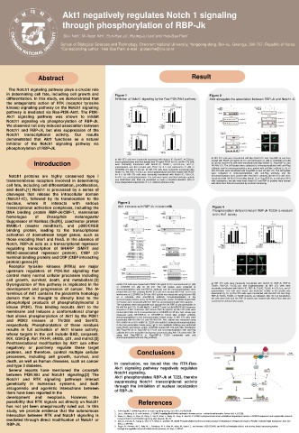
Akt1 negatively regulates Notch 1 signaling
through phosphorylation of RBP-Jk
School of Biological Sciences and Technology, Chonnam National University, Yongbong-dong, Buk-ku, Gwangju, 500-757, Republic of Korea
*Corresponding author : Hee-Sae Park, e-mail : proteome@jnu.ac.kr
Abstract Result
The Notch1 signaling pathway plays a crucial role
in determining cell fate, including cell growth and Figure 1 Figure 2
differentiation. In this study, we demonstrated that Inhibition of Notch1 signaling by the Ras-PI3K/Akt1 pathway Akt1 abrogates the association between RBP-Jk and Notch1-IC
the antagonistic action of RTK (receptor tyrosine
kinase) signaling pathway on the Notch1 signaling
pathway is mediated via Ras-PI3K-Akt1. The PI3K-
Akt1 signaling pathway was shown to inhibit
Notch1 signaling via phosphorylation of RBP-Jk.
We observed not only reduced association between
Notch1 and RBP-Jk, but also suppression of the
Notch1 transcriptional activity. Our results
demonstrated that Akt1 functions as a natural
inhibitor of the Notch1 signaling pathway via
phosphorylation of RBP-Jk.
a) NIH 3T3 cells were transfected with Myc-Notch1-IC and Flag-RBP-Jk and then
a) NIH 3T3 cells were transiently transfected with Notch1-IC, RasN17, 4×CSLLuc, treated with PDGF (10 ng/ml) for 8 h and wortmannin (1 μM) or LY294002 (10 μM)
Introduction and β-galactosidase and then treated with 10 ng/ml PDGF for 8 h. b) NIH 3T3 cells for 30 min. b) NIH 3T3 cells were transfected with Myc-Notch1-IC, Flag-RBP-Jk, and
were transiently transfected with Notch1-IC, RasN17, 4×CSL-Luc, and β-
galactosidase and then treated with PDGF for 8 h and wortmannin (1 μM) or HA-Akt1-CA. The cell lysates were subjected to immunoprecipitation with anti-Flag
antibody, and the immunoprecipitates were probed with anti-Myc antibody (a, b). c)
LY294002 (10 μM) for 30 min. c) NIH 3T3 cells were transiently transfected with NIH 3T3 cells were transfected with Flag-RBP-Jk and HA-Akt1-CA. The cell lysates
Notch1-IC, Akt1-DN, 4×CSL-Luc, and β-galactosidase and then treated with PDGF were subjected to immunoprecipitation with anti-Flag antibody, and the
Notch1 proteins are highly conserved type I for 8 h. d) NIH 3T3 cells were transiently transfected with Notch1-IC, Akt1-CA, immunoprecipitates were probed with Anti-SMRT antibody. d) NIH 3T3 cells were
4×CSL-Luc, and β-galactosidase. The cells were lysed, and their luciferase activity
transmembrane receptors involved in determining was determined (ad). Data are expressed as mean ± standard deviation (SD) of transfected with HA-Akt1-CA and Myc-Notch1-IC or Flag-RBP-Jk, as indicated; 48 h
after transfection, the cells were fixed; Notch1-IC and RBP-Jk proteins were stained
three independent experiments; R.L.U., relative luciferase units.
cell fate, including cell differentiation, proliferation, with Alexa Fluor 488 and examined by confocal microscopy.
and death.[1] Notch1 is processed by a series of
cleavages that release the intracellular domain
(Notch1-IC), followed by its translocation to the
nucleus, where it interacts with various Figure 3
transcriptional activation complexes, including the Akt1 interacts with RBP-Jk in intact cells Figure 4
DNA binding protein RBP-Jk/CBF-1, mammalian Phosphorylation deficient mutant RBP-Jk T223A is resistant
to the Akt1 activity
homologue of Drosophila melanogaster
Suppressor of Hairless [Su(H)], coactivator protein
MAML-1 (master mindlike1), and p300/CREB
binding protein, leading to the transcriptional
activation of downstream target genes, such as
those encoding Hes1 and Hes5. In the absence of
Notch, RBP-Jk acts as a transcriptional repressor
regulating transcription of SHARP (SMRT and
HDAC-associated repressor protein), CtBP (C-
terminal binding protein) and CtIP (CtBP interacting
protein) genes.[4]
Receptor tyrosine kinases (RTKs) are major
upstream regulators of PI3K/Akt signaling that
control many normal cellular processes including
cell growth, survival, death, and metabolism.[2]
Dysregulation of this pathway is implicated in the a) NIH 3T3 cells were treated with PDGF (10 ng/ml) for 8 h and wortmannin (1 μM) a) NIH 3T3 cells were transiently transfected with Notch1-IC, RBP-Jk, RBP-Jk
T223A, Akt1-CA, 4×CSL-Luc, and β-galactosidase. b) NIH 3T3 cells were
development and progression of cancer. The N- or LY294002 (10 μM) for 30 min. The cell lysates were subjected to transiently transfected with Notch1-IC, RBP-Jk, T223E, T223D, 4×CSL-Luc, and β-
immunoprecipitation with anti-RBP-Jk antibody, and the immunoprecipitates were
galactosidase. The cells were lysed, and luciferase activity in the lysates was
terminus of Akt1 contains the pleckstrin homology probed with anti-Akt1 antibody. b) NIH 3T3 cells were treated with PDGF for 8 h and determined (a, b). c) NIH 3T3 cells were transfected with HA-Akt1-CA and Flag-
wortmannin or LY294002 for 30 min. Akt1 activity was measured using histone H2B
domain that is thought to directly bind to the as a substrate after anti-RBP-Jk antibody immunoprecipitation in the RBP-Jk T223A, T223D, or T223E mutants, as indicated. After 48 h of transfection,
the cells were fixed and the RBP-Jk protein was stained with Alexa Fluor 488 and
immunocomplex kinase assay. c) PDGF, wortmannin, and/or LY294002 treated NIH
phospholipid products of phosphatidylinositol 3 3T3 lysates were incubated with GST-RBP-Jk and precipitated with GSH-agarose. examined by confocal microscopy.
kinase (PI3K). This binding recruits Akt1 to the The complexes were resuspended and assayed for the RBP-Jk phosphorylation in
50 mM Tris (pH 7.5), 10 mM MgCl2, 1 mM DTT, and 2 μCi[γ32P]ATP. d) Consensus
membrane and induces a conformational change sequence of Akt1 substrates and various RBP-Jk species. e) NIH 3T3 cells were
treated with PDGF for 8 h and wortmannin or LY294002 for 30 min. Akt1 activity was
that allows phosphorylation of Akt1 by the PDK1 measured using GST-RBP-Jk or GST-RBP-Jk T223A after antiAkt1 antibody
immunoprecipitation in the immunocomplex kinase assay. f) NIH 3T3 cells were
and PDK2 kinases at Thr308 and Ser473, transfected with HA-Akt1-CA and HA-Akt1-DN. Akt1 activity was measured using
GST-mRBP-Jk or GST-mRBP-Jk T223A after antiAkt1 antibody immunoprecipitation
respectively. Phosphorylation of these residues in the immunocomplex kinase assay. g) In vivo metabolic labeling was performed
results in full activation of Akt1 kinase activity, using PDGF, wortmannin, and/or LY294002 treated NIH 3T3 cells after transfection
with Flag-RBP-Jk or Flag-RBP-Jk T223A and immunoprecipitation with anti-Flag
whose targets in the cell include BAD, caspase9, antibody. h) In vivo metabolic labeling was performed using Akt1-CA and/or Akt1-DN
with
cells
T223A
transfected
Flag-RBP-Jk
or
and
Flag-RBP-Jk
along
IKK, GSK3-β, Raf, FKHR, eNOS, p21, and mdm2.[5] immunoprecipitated with anti-Flag antibody.
Posttranslational modification by Akt1 can either
negatively or positively regulate these target
proteins, and therefore, control multiple cellular Conclusions
processes, including cell growth, survival, and
death, as well as human diseases, such as cancer
and type 2 diabetes. In conclusion, we found that the RTK-Ras-
Several reports have mentioned the crosstalk Akt1 signaling pathway negatively regulates
between PI3K/Akt and Notch1 signaling.[3] The Notch1 signaling.
Notch1 and RTK signaling pathways interact Akt1 phosphorylates RBP-Jk at T223, thereby
genetically in numerous systems, and both suppressing Notch1 transcriptional activity
antagonistic and agonistic interactions between through the inhibition of nuclear localization
them have been reported in the of RBP-Jk.
development and neoplasia. However, the
possibility that RTK signals act directly on Notch1 References
has not yet been unequivocally ruled out. In this
study, we provide evidence that the autonomous 1. Schweisguth, F. (2004) Regulation of notch signaling activity, Curr. Biol., 14, R129138.
interaction between RTK and Notch1 signaling is 2. Luo, J., Manning, B. D., and Cantley, L. C. (2003) Targeting the PI3KAkt pathway in human cancer: rationale and promise, Cancer Cell, 4, 257262.
3. Wang, H., Cheng, H., Shao, Q., Dong, Z., Xie, Q., Zhao, L., Wang, Q., Kong, B., and Qu, X. (2014) Leptinpromoted human extravillous trophoblast invasion is MMP14 dependent and requires the crosstalk
mediated through direct modification of Notch1 or 4. Gao, D., Inuzuka, H., Tseng, A., Chin, R. Y., Toker, A., and Wei, W. (2009) Phosphorylation by Akt1 promotes cytoplasmic localization of Skp2 and impairs APCCdh1 mediated Skp2 destruction, Nat. Cell
between Notch1 and PI3K/Akt signaling, Biol. Reprod., 90, 78.
RBP-Jk. Biol., 11, 397408.
5. Riggio, M., Perrone, M. C., Polo, M. L., Rodriguez, M. J., May, M., Abba, M., Lanari, C., and Novaro, V. (2017) AKT1 and AKT2 isoforms play distinct roles during breast cancer progression
through the regulation of specific downstream proteins, Sci. Rep., 7, 44244.

