


Page 33 - ebook
P. 33
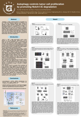
Autophagy controls tumor cell proliferation
by promoting Notch1-IC degradation
School of Biological Sciences and Technology, Chonnam National University, Yongbong-dong, Buk-ku, Gwangju, 500-757, Republic of Korea
*Corresponding author : Hee-Sae Park, e-mail : proteome@jnu.ac.kr
Abstract Result
Autophagy is a highly conserved mechanism that degrades long-
lived proteins and dysfunctional organelles, and contributes to cell
fate. In this study, autophagy attenuates Notch1 signaling by Figure 1 Figure 2
degrading the Notch1 intracellular domain (Notch1-IC). Nutrient- Autophagy attenuates Notch1 signaling Autophagy induces Notch1-IC degradation
deprivation promotes Notch1-IC phosphorylation by MEKK1 and
phosphorylated Notch1-IC is recognized by Fbw7 E3 ligase. The
ubiquitination of Notch1-IC by Fbw7 is essential for the interaction
between Notch1-IC and p62 and for the formation of aggregates.
Inhibition of Notch1 signaling prevents the transformation of breast
cancer cells, tumor progression, and metastasis. The expression of
Notch1 and p62 is inversely correlated with Beclin1 expression in
human breast cancer patients. These results show that autophagy
inhibits Notch1 signaling by promoting Notch1-IC degradation and
therefore plays a role in tumor suppression. .
Introduction (A) Rapamycin (Rap) treatment and nutrient deprivation attenuate the Notch1-IC transcriptional activity.
HEK293 cells were transfected with the 4xCSL-Luc, together with pcDNA3 or Myc-Notch1-IC plasmids. After
48 h of transfection, the cells were treated with 2 μM rap, 10 mM 3-MA, or nutrient deprivation (ND) for 6 h,
as indicated, and analyzed for Notch1-IC transcriptional activity (fold induction). (B) Nutrient deprivation
reduces Notch1 target gene mRNA expression. HEK293 cells with pcDNA3 or Myc-Notch1-IC plasmids were (A) Notch1-IC is degraded by autophagy. HEK293 cells were treated with 2 μM rap or nutrient-deprivation
starved for 4 hr. After RNA extraction and cDNA synthesis, quantitative RT-PCR was performed. (C) medium. Each condition was reversed by treating 10 mM 3-MA, autophagy inhibitor. The cell lysates were
Notch1 is a type 1 single transmembrane receptor protein Knockdown of autophagy mediator LC3, Beclin1, and p62 induce Notch1-IC transcriptional activity. HEK293 subjected to immunoblotting. (B) Half-life of Notch1-IC was extended in Atg5−/− and Atg7−/− MEFs. WT and
important for cell fate specification, differentiation in various cells with pcDNA3 or Myc-Notch1-IC plasmids were transfected with shCon, shBeclin1, or shLC3 Atg5−/− and Atg7−/− MEFs were starved for the indicated durations and subjected to immunoblotting.
respectively. After transfection, the cells were analyzed for Notch1-IC transcriptional activity. (D) Knockdown
Notch1-IC levels were quantified by ImageJ (Right panel). (C) Atg5−/− MEFs with reintroduction of Atg5
systems and neuronal development such as neurogenesis and of Atg5 enhanced Notch1 signaling. Atg5+/+ and Atg5−/− MEFs were transfected with the 4xCSL-Luc, promotes the Notch1-IC degradation. Atg5−/− MEFs with pcDNA3 or GFP-Atg5 were starved for the
together with pcDNA3 or Myc-Notch1-IC. After transfection, the cells were analyzed for Notch1-IC
indicated durations and subjected to immunoblotting. (D) The continued degradation of Notch1-IC in Atg5−/−
neural stem cell maintenance.[1] Notch1 signaling is aberrantly transcriptional activity. The relative luciferase activities were normalized with β-galactosidase activity. Data MEFs is proteasome-dependent. WT and Atg5−/− MEFs were starved for 2, 4, or 8 h with 1 μM MG132 or
represent the mean ± SD from independent experiments performed in triplicate. ‘ND’ means nutrient-
activated in breast cancer, and increased expression of the Notch1 deprivation. **p < 0.01; ***p < 0.001. 100 μM BFA1 to inhibit the different degradation systems and lysed for immunoblotting analysis. LC3 and
p62 were used as autophagy markers. Results are representative of at least 3 independent experiments. ‘Nut’
intracellular domain (Notch1-IC) is associated with low survival rates means nutrient-rich. β-actin was used as a loading control.
in various cancers, including breast cancer.[2] Proliferation of cells
derived from these cancers can be suppressed by pharmacological
inhibition of Notch1. Therefore, preventing the generation of Notch1-
IC is a potential strategy for treating various cancers. Figure 3 Figure 4
Genetic analysis of Drosophila melanogaster revealed a possible Nutrient-deprivation promotes the interaction between Notch1-IC Ubiquitination of Notch1-IC under nutrient-deprivation condition
link between autophagy and the Notch1 signaling pathway involved and LC3, which is facilitated by the interaction between Notch1- by Fbw7 is critical for Notch1-IC-p62 interaction and degradation
in cell fate determination.[3] The mechanistic target of rapamycin IC with p62. via autophagy.
(mTOR), a negative regulator of autophagy, activates Notch1
signaling.[5] The lack of autophagy triggers precocious activation of
Notch1 signaling during Drosophila oogenesis, suggesting that
autophagy suppresses Notch1 signaling. However, the relationship
between autophagy and Notch1 signaling in tumorigenesis and the
precise regulatory mechanism is not well known. Many reports
found that defective autophagy causes various cancers. Beclin1 or
UVRAG are monoallelically deleted in a high percentage of human
breast and colon cancers respectively.
Atg5, a component of the ubiquitin-like protein conjugation systems,
and Beclin1 have tumor suppressor effects in mouse xenograft
models. It is clear these autophagy-related genes are involved in the
regulation of tumorigenesis but it is not clear whether autophagy
attenuates the tumorigenesis through the inhibition of oncogenic
signal transduction.
In this study, we evaluated the crosstalk between autophagy and
Notch1 signaling during tumorigenesis. We discovered that
autophagic stimuli induced MEKK1 to phosphorylate the T2512
residue of Notch1-IC enabling its ubiquitination and degradation by
Fbw7 ubiquitin ligase.[4] We also found that the expression of (A) Nutrient-deprivation promotes ubiquitination of Notch1-IC. HEK293 cells transfected with indicated
plasmids were cultured with BFA1 under nutrient-rich or nutrient-deprivation conditions. Cell lysates were
Notch1 and Beclin1 protein in tissues of patients with breast cancer (A) Localization of Notch1-IC and LC3 under nutrient-rich and nutrient-deprivation conditions. HEK293 cells precipitated by Ni-NTA and were subjected to immunoblotting using anti-Myc antibody. (B) Nutrient-
with GFP-LC3 were cultured with 0.1 μM BFA1 under nutrient-rich and nutrient-deprivation conditions. The
were negatively correlated. Notch1 inhibition significantly decreased cells were stained with anti-Notch1-IC V1744 antibody and ToPro3. (B) Notch1-IC binds to LC3. deprivation promotes the interaction of Notch1-IC with Fbw7. HEK293 cells transfected with indicated
plasmids were cultured with BFA1 under nutrient-rich or nutrient-deprivation conditions. Immunoprecipitates
Immunoprecipitates by anti-IgG control or anti-LC3 antibody from HEK293 cells cultured with BFA1 under
growth, invasion, and tumorigenic activity of Beclin1 knockdown nutrient-rich or nutrient-deprivation conditions were subjected to immunoblotting using anti-Notch1 antibody. by anti-Myc antibody from the cells were subjected to immunoblotting using anti-Flag antibody. (C) Under
nutrient-deprivation conditions, the ubiquitination of Notch1-IC is Fbw7-dependent. HEK293 cells transfected
cells. These data suggested that autophagy-induced MEKK1- (C) p62 promotes the interaction of Notch1-IC with LC3. Immunoprecipitates by anti-GFP antibody from with indicated plasmids were cultured with BFA1 under nutrient-deprivation conditions. Immunoprecipitates
HEK293 cells transfected with indicated plasmids were subjected to immunoblotting using anti-Myc antibody.
by anti-Myc antibody from the cells were subjected to immunoblotting using anti-HA antibody. (D)
mediated phosphorylation of Notch1-IC at the T2512 residue plays (D) Notch1-IC binds to p62. Immunoprecipitates by anti-IgG control or anti-p62 antibody from HEK293 cells Ubiquitination of Notch1-IC is decreased by shFbw7. HEK293 cells with shCon or shFbw7 were transfected
cultured with BFA1 under nutrient-rich or nutrient-deprivation conditions were subjected to immunoblotting
an important role in cancer prevention and could be a promising using anti-Notch1 antibody. (E) Knockdown of p62 decreases the interaction of Notch1-IC with LC3. HEK293 with indicated plasmids. The cells were cultured with BFA1 under nutrient-rich or nutrient-deprivation
conditions. Immunoprecipitates by anti-HA antibody from the cells were subjected to immunoblotting using
cells with shCon or shp62 were cultured with BFA1 under nutrient-rich or nutrient-deprivation conditions.
strategy to prevent cancer progression. Immunoprecipitates by anti-IgG control or anti-LC3 antibody from the cells were subjected to immunoblotting anti-Myc antibody. (E) F-box domain in Fbw7 is required for Notch1-IC and p62 interaction.
using anti- Notch1 antibody. (F) p62 is important for Notch1-IC aggregation and translocalization to Immunoprecipitates by anti-Myc antibody from HEK293 cells transfected with indicated plasmids were
autophagosome. HEK293 cells were transfected with shCon or shp62. The cells were cultured with BFA1 subjected to immunoblotting using anti-HA antibody. (F) Knockdown of Fbw7 decreases the interaction of
under nutrient-rich medium or nutrient-deprivation conditions and stained with anti-p62, anti-Notch1-IC Notch1-IC with p62. HEK293 cells with shCon or shFbw7 were cultured with BFA1 under nutrient-rich or
nutrient-deprivation conditions. Immunoprecipitates by anti-IgG control or anti-p62 antibody from the cells
V1744 antibodies, and ToPro3. (G) Half-life of Notch1-IC was extended by knockdown of p62. HEK293 cells were subjected to immunoblotting using anti-Notch1 antibody. (G) Polyubiquitination is required for Notch1-IC
Conclusions with shCon or shp62 were starved for the indicated durations and subjected to immunoblotting. All images and p62 interaction. HEK293 cells with His-Ub or His-Ub-7KR were cultured with BFA1 under nutrient-rich or
were confocal images of optical slice thickness ~1 μm. Scale bars represent 25 μm. Results are
nutrient-deprivation conditions. Immunoprecipitates by anti-IgG control or anti-p62 antibody from the cells
representative of at least 3 independent experiments. ***p < 0.001; ns: not significant (p > 0.05).
were subjected to immunoblotting using anti-Notch1 antibody. Results are representative of at least 3
independent experiments.
• nutrient-deprivation condition, MEKK1 phosphorylates the
Notch1-IC T2512 residue enabling Fbw7 to recognize and
ubiquitinate the phosphorylated Notch1-IC. Figure 5
Notch1-IC phosphorylation by MEKK1 is critical for the regulation Figure 6
• autophagy promotes the degradation of the Notch1 intracellular of Notch1-IC stability during nutrient-deprivation. Negative correlation between Notch1-IC and autophagy in breast
domain via the phosphorylation of the T2512 residue by MEKK1, cancer
and therefore suppresses tumor cell growth and migration
• autophagy decreases Notch1 signaling-dependent tumorigenesis.
(A) Nutrient-deprivation promotes ubiquitination of Notch1-IC. HEK293 cells transfected with indicated
plasmids were cultured with BFA1 under nutrient-rich or nutrient-deprivation conditions. Cell lysates were (A) Notch1-IC protein stability is increased by knockdown of Beclin1. HEK293 cells were transfected with
precipitated by Ni-NTA and were subjected to immunoblotting using anti-Myc antibody. (B) Nutrient- shBeclin1 in dose-dependent manner and subjected to immunoblotting using anti-Notch1-IC, anti-Beclin1,
deprivation promotes the interaction of Notch1-IC with Fbw7. HEK293 cells transfected with indicated and anti-β-actin antibodies. (B–E) MDA-MB-231 cells stably expressing shCon or shBeclin1 were treated
plasmids were cultured with BFA1 under nutrient-rich or nutrient-deprivation conditions. Immunoprecipitates with or without 2 μM DAPT. B. Knockdown of Beclin1 induces wound-healing migration ability. The wound
by anti-Myc antibody from the cells were subjected to immunoblotting using anti-Flag antibody. (C) Under closure was quantified for the indicated durations and was analyzed by measuring the width of the remaining
nutrient-deprivation conditions, the ubiquitination of Notch1-IC is Fbw7-dependent. HEK293 cells transfected unmigrated area. Wound area is plotted (right). C. Knockdown of Beclin1 induces cell migration ability. The
with indicated plasmids were cultured with BFA1 under nutrient-deprivation conditions. Immunoprecipitates cells were seeded onto fibronectin-coated Transwell inserts. The migrating cells were stained by DAPI and
by anti-Myc antibody from the cells were subjected to immunoblotting using anti-HA antibody. (D) quantified by counting. The number of migrated cells is plotted (right). D. Knockdown of Beclin1 induces cell
Ubiquitination of Notch1-IC is decreased by shFbw7. HEK293 cells with shCon or shFbw7 were transfected invasion ability. The cells were seeded onto a Matrigel invasion chamber. The invading cells were stained by
with indicated plasmids. The cells were cultured with BFA1 under nutrient-rich or nutrient-deprivation DAPI and quantified by counting. The number of migrated cells is plotted (right). E. Knockdown of Beclin1
References conditions. Immunoprecipitates by anti-HA antibody from the cells were subjected to immunoblotting using induces the ability of the cells to form anchorage-dependent colonies. The cells were seeded onto 24 well
anti-Myc antibody. (E) F-box domain in Fbw7 is required for Notch1-IC and p62 interaction.
plates at a density of 100 cells per well and were incubated for 10 days. The cell colonies were stained with
Immunoprecipitates by anti-Myc antibody from HEK293 cells transfected with indicated plasmids were
subjected to immunoblotting using anti-HA antibody. (F) Knockdown of Fbw7 decreases the interaction of crystal violet. The graph shows the number of colonies formed by each cell. (F) Knockdown of Beclin1
induces the ability of the cells to form anchorage-independent colonies. The cells were plated at a density of
Notch1-IC with p62. HEK293 cells with shCon or shFbw7 were cultured with BFA1 under nutrient-rich or 5 × 10³ cells in a top medium containing 0.3% agarose and were incubated for 10 days. The cell colonies
1. Artavanis-Tsakonas S, Rand MD, Lake RJ. Notch signaling: cell fate control and signal integration in development. Science. nutrient-deprivation conditions. Immunoprecipitates by anti-IgG control or anti-p62 antibody from the cells were stained with crystal violet. The graph shows the number of colonies formed by each cell. (G) and (H)
1999; 284:770–776. were subjected to immunoblotting using anti-Notch1 antibody. (G) Polyubiquitination is required for Notch1-IC Negative correlation of Notch1-IC and autophagy in human breast cancer samples versus normal tissues. (G)
2. Pece S, Serresi M, Santolini E, Capra M, Hulleman E, Galimberti V, Zurrida S, Maisonneuve P, Viale G, Di Fiore PP. Loss and p62 interaction. HEK293 cells with His-Ub or His-Ub-7KR were cultured with BFA1 under nutrient-rich or Lysates of paired human normal and breast cancer tissues were subjected to immunoblotting using anti-
of negative regulation by Numb over Notch is relevant to human breast carcinogenesis. J Cell Biol. 2004; 167:215–21. doi: nutrient-deprivation conditions. Immunoprecipitates by anti-IgG control or anti-p62 antibody from the cells Notch1, anti-Beclin1, anti-p62, and anti-β-actin antibodies. (H) Ranking plot of each protein in normal and
10.1083/jcb.200406140. were subjected to immunoblotting using anti-Notch1 antibody. Results are representative of at least 3 breast tumor tissues. The difference in Notch1-IC, Beclin1 and p62 between normal and tumor tissue were
3. Gao C, Cao W, Bao L, Zuo W, Xie G, Cai T, Fu W, Zhang J, Wu W, Zhang X, Chen YG. Autophagy negatively regulates independent experiments. examined by Wilcoxon Signed-Rank test. Results are representative of at least 3 independent experiments.
Wnt signalling by promoting Dishevelled degradation. Nat Cell Biol. 2010; 12:781–90. doi: 10.1038/ncb2082. Data represent means ± SD from independent experiments performed in triplicate. *p < 0.05; **p < 0.01; ***p
4. O’Neil J, Grim J, Strack P, Rao S, Tibbitts D, Winter C, Hardwick J, Welcker M, Meijerink JP, Pieters R, Draetta G, Sears R, < 0.001.
Clurman BE, et al. FBW7 mutations in leukemic cells mediate NOTCH pathway activation and resistance to gamma-
secretase inhibitors. J Exp Med. 2007; 204:1813– 24. doi: 10.1084/jem.20070876.
5. Chan SM, Weng AP, Tibshirani R, Aster JC, Utz PJ. Notch signals positively regulate activity of the mTOR pathway in T-cell
acute lymphoblastic leukemia. Blood. 2007; 110:278–286. doi: 10.1182/blood-2006-08-039883.

