Page 13 - ebook
P. 13

Cancer patient tissueoid with self-homing nano-targeting of


                                                                                                                                                                                               metabolic inhibitor


                                                                                                                                                                                                                                                                                                                                                                                           1
                                                                                                                                                                                                                              2*
                                                                                                                                                                           2*
                                                                                                                                                                                                                                                                                                                                                                                                                                                2
                                                                                                         1*
                                                                                                                                                                                                                                                                                   1
                                                                                                                                                                                                                                                                                                                                       1
                                                                Hyo-Jin Yoon , Young Shin Chung , Yong Jae Lee , Seung Eun Yu , Sewoom Baek , Hye-Seon Kim , Sang Wun Kim ,
                                                                                                                                                                                                              2†
                                                                                                                                                                   Jung-Yun Lee , Sunghoon Kim , and Hak-Joon Sung                                                                                                                          1†
                                                                                                                                                                                                                                                                     2†


                                                                                                                    1 Department of Medical Engineering, Yonsei University College of Medicine, Seoul 03722, Republic of Korea.


                                                     2 Institute of Women's Life Medical Science, Department of Obstetrics and Gynecology, Yonsei University College of Medicine, Seoul 03722, Republic of Korea.


                                                                                                    ABSTRACT                                                                                                                                                                                                                                      INTRODUCTION


          The current paradigm of cancer medicine focuses on patient- and/or cancer-specific                                                                                                                                                                          Recent advances in biotechnologies have been integrated with precision medicine. As a


          treatments, which has led to continuous progress in the development of patient                                                                                                                                                                              major outcome, the paradigm of anti-cancer drug treatment has become to focus on


          representatives (e.g., organoids) and cancer-targeting carriers for drug screening. As                                                                                                                                                                      patient-specific selection and cancer-specific targeting                                                                                                [1, 2]   . A common goal of current


          breakthrough concepts, i) living cancer tissues convey intact profiles of patient-specific                                                                                                                                                                  outstanding technologies is to expedite clinical success by cross-validating the safety,


          microenvironmental signatures. ii) The growth mechanisms of cancer mass with intense                                                                                                                                                                        efficacy, and efficiency of anti-cancer drugs among in vitro, in vivo, and clinical studies.


          cell-cell interactions can be harnessed to develop self-homing nano-targeting by using                                                                                                                                                                      To this end, extensive efforts have been made to develop artificial models (e.g., cell and


          cancer cell-derived nanovesicles (CaNVs). Hence, we developed a tissueoid model of                                                                                                                                                                          organoid) of patient representatives [3, 4] for drug screening as a potential platform for


          ovarian cancer (OC) by culturing OC patient tissues in a 3D gel chip, whose                                                                                                                                                                                 cross-validation with in vivo responses through implantation [5, 6]. the breakthrough


          microchannel networks enable perfusion to maintain the tissue viability. A novel model                                                                                                                                                                      strategy of self-homing nano-targeting can be applied. The reciprocal interaction of cell


          of systemic cancer responses was approached by xenografting OC tissueoids into                                                                                                                                                                              membranes is a key mediator of cancer cell-cell interactions, whose function can be


          ischaemic hindlimbs in nude mice. CaNVs were produced to carry general                                                                                                                                                                                      potentiated using cancer cell-derived nanovesicles (CaNV), owing to i) the same


          chemotherapeutics or new drugs under pre/clinical studies that target the BRCA                                                                                                                                                                              membrane characteristics as target cancer cells; ii) inanimation to prevent living cell-


          mutation or energy metabolism, thereby increasing the test scope. This pioneer study                                                                                                                                                                        mediated side effects; iii) effective systemic delivery similar to exosomes [7-9], as


          cross-validates drug responses from the OC clinic, tissueoid, and animal model by                                                                                                                                                                           revealed by another group [10]; and iv) more efficient mass production and purification


          demonstrating the alignment of results in drug type-specific efficiency, BRCA mutation-                                                                                                                                                                     [11, 12] compared to exosomes [13-16]. Moreover, the loading of anti-cancer drugs into


          dependent drug efficiency, and metabolism inhibition-based anti-cancer effects. Hence,                                                                                                                                                                      CaNV represents a “Trojan horse” strategy to inhibit cancer action synergistically with the


          this study provides a directional foundation to accelerate the discovery of patient-                                                                                                                                                                        self-homing nano-targeting approach. Therefore, this study introduces the novel concept


          specific drugs with CaNV application towards future precision medicine.                                                                                                                                                                                     of an implantable OC tissueoid capable of culturing OC patient tissues in vitro and in vivo.


                                                                                                                                                                                                                                                                      Drug type-specific efficiency, BRCA mutation-dependent drug efficiency, and metabolism


                                                                                                                                                                                                                                                                      inhibition-based anti-cancer effects were tested. CaNVs were produced to deliver general
                                                                                              OBJECTIVES                                                                                                                                                              chemotherapeutics, as well as the two aforementioned drugs following the concept of


                                                                                                                                                                                                                                                                      Trojan horse. Based on more than 100 patient records, the cross-validation among in


           To demonstrate breakthrough concepts including the OC tissueoid, CaNV, new drugs, in vivo                                                                                                                                                                  vitro, in vivo, and clinical studies were carried out as the first step of a paradigm shift in


         model of systemic cancer response, and to cross-check with clinical outcomes.                                                                                                                                                                                the field of new drug development.


                                                                                                                                                                                                                                        RESULTS


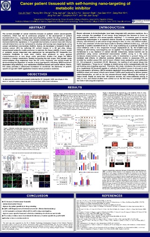
                                                                                                                                                                                                                                                                                                                                                               Figure 3. Xenografting OC tissueoid as a new systemic model to cross-
                                                                                                                                                                                       Figure 2. Cancer cell derived-nanovesicles (CaNVs) for self-homing nano-                                                                                                validate in vivo anti-cancer effects of MB-Drug. (a) At week seven post OC

                                                                                                                                                                                       targeting of MB-Drug. (a) CaNV is produced by serial filtering of OC cells with                                                                                         tissueoid xenografting into ischemic hindlimbs of nude mice, tumor growth over 200

                                                                                                                                                                                       decreasing pore sizes and loading with the MB-Drug (i.e., a combination of                                                                                              mm in volume was established. Then, test groups (no treatment– PBS vehicle
                                                                                                                                                                                                                                                                                                                                                                     3
                                                                                                                                                                                       gossypol             and         phenformin)               by       electroporation.                CaNVs+/–MB-Drug                         are                         control, CaNVs, CaNVs+MB-Drug, MB-Drug) were injected intraperitoneally every

                                                                                                                                                                                       characterized for (b) size by DLS and (c) morphologies by TEM. (d) The loading                                                                                          week until sacrifice. Tumor tissues were harvested at week 14. Anti-cancer effects

                                                                                                                                                                                       of MB-Drug into CaNV was verified by analysing each component with MALDI-                                                                                               of test groups were determined in terms of (b) optical size imaging with a ruler, (c)

                                                                                                                                                                                       TOF/TOF. (e) Cancer-specific cytotoxic effects of test groups were determined by                                                                                        tumor weight (gram: g), and (d) quantitative analyses of progressive changes in

                                                                                                                                                                                       analysing the viability of normal vs. OC tissueoid with confocal image analysis,                                                                                        tumor size (mm ). (e) The mitotic index, proliferation (Ki67), and Notch-1 factor
                                                                                                                                                                                                                                                                                                                                                                                            3
                                                                                                                                                                                                                                                                            +
                                                                                                                                                                                       which is further supported by (f) OC cell (PAX8 )-specific targeting and uptake of                                                                                      expression (cleaved Notch-1 and HES-1) were analysed quantitatively with
                                                                                                                                                                                       CaNV+MB-Drug in mixture culture with normal ovarian cells by phase contrast                                                                                             immune-histological staining (arrows in H&E images: apoptotic cells), (f) followed by

                                                                                                                                                                                       and immunofluorescence imaging. Data = mean ± S.E.M (n  3). *p < 0.05, **p                                                                                             Western blot analyses of Notch signaling proteins for crosschecking. Data = mean

                                                                                                                                                                                       < 0.01, and ***p < 0.001 versus no treatment or between lined groups.                                                                                                   ± S.E.M (n = 6 mice). *p < 0.05, **p < 0.01, and ***p < 0.001 versus No treat or


                                                                                                                                                                                                                                                                                                                                                               between lined groups.


             Figure 1. Clinically matched drug responses of ovarian cancer (OC) tissueoid

             with introduction of anti-cancer metabolic (MB)-Drug. (a) The tissueoid is

             defined as a culture of patient tissues in a 3D chip under perfusion of media (+/–

             drug) into microchannel (vascular) networks at a flow rate of 20 μL/min. (b) OC

             marker expression (PAX8 and p53) and histology of tissueoids were matched with

             those of tissue samples from high-grade serious OC patients in-clinic, analysed by

             optical examination with immunohistology and H&E staining. (c) The gelatin gel

             allows the progressive cell invasion from OC patient tissues during 28-day culture

             post embedding into the gel, as observed by optical imaging. (d) The viability of the

             OC tissueoid was maintained during 30 days of perfusion culturing, as determined

             by confocal imaging. (e) Gossypol and phenformin were used in combination as an

             anti-cancer energy metabolism (MB)-Drug. (f) To represent patient tissue [+/- BRCA

             mutation (mut)], drug type-dependent viabilities of OC tissueoids were validated

             under treatments with paclitaxel (PTX) + carboplatin (CBP), olaparib (Ola), or MB-

             Drug by confocal imaging analysis. Data = mean ± S.E.M (n 3). *p < 0.05, **p

             < 0.01, and ***p < 0.001 versus no treatment or between lined groups.


                                                                                                                                                                                                                                                                                                                                                             Figure 4. Aligned drug responses between OC tissueoid and patient tissue in-

                                                                                                                                                                                                                                                                                                                                                             clinic. (a) During clinical progress in OC treatment, tissues were obtained from

                                                                                                                                                                                                                                                                                                                                                             diagnosis (*) and surgery in-between PTX+CBP (**) treatment for the drug-first

                                                                                                                                                                                                                                                                                                                                                             group (top) or surgery before the first PTX+CBP (***) administration for the surgery-

                                                                                                                                                                                                                                                                                                                                                             first group (bottom). (b) In alignment with the tissuoid responses, the first PTX+CBP

                                                                                                                                                                                                                                                                                                                                                             therapy exhibits anti-cancer effects (>50 %) on BRCA-WT and BRCA-mut groups (n

                                                                                                                                                                                                                                                                                                                                                             = 82). However, significant recurrence rates (>30 %) indicate the need to use

                                                                                                                                                                                                                                                                                                                                                             another drug. (c) Hence, Ola was administered to each group as a maintenance

                                                                                                                                                                                                                                                                                                                                                             therapy after PTX+CBP, resulting in a more effective tumor reduction (14.3 %) in the

                                                                                                                                                                                                                                                                                                                                                             BRCA-mut group compared to the BRCA-WT group (29.4 %). (d) As an example of

                                                                                                                                                                                                                                                                                                                                                             alignment with the OC tissueoid response, a clinical case is shown post first

                                                                                                                                                                                                                                                                                                                                                             PTX+CBP therapy. The effective drug response is evidenced by the significantly

                                                                                                                                                                                                                                                                                                                                                             lowered level of the standard tumor marker (CA 125: 13.7 U/mL) below the normal

                                                                                                                                                                                                                                                                                                                                                             limit (35 U/mL), which was verified by PET/CT imaging. (Red circle: tumor mass).


                                                                                            CONCLUSION                                                                                                                                                                                                                                                      REFERENCES


                                                                                                                                                                                                                                                                      [1] Z. Hu, P. A. Ott, C. J. Wu. Nat Rev Immunol. 2018, 18, 168-182.
        ● OC tissueoid (Ovarian cancer tissueoid)                                                                                                                                                                                                                     [2] L. Scheetz, K. S. Park, Q. Li, P. R. Lowenstein, M. G. Castro, A. Schwendeman, J. J. Moon. Nat Biomed Eng. 2019, 3, 768-782.


        - culture intact patient tissues                                                                                                                                                                                                                              [3] S. N. Bhatia, D. E. Ingber. Nat Biotechnol. 2014, 32, 760-772.
                                                                                                                                                                                                                                                                      [4] K. Ronaldson-Bouchard, G. Vunjak-Novakovic. Cell Stem Cell. 2018, 22, 310-324.

        - improve the patient-specificity for drug screening                                                                                                                                                                                                          [5] M. M. Laronda, A. L. Rutz, S. Xiao, K. A. Whelan, F. E. Duncan, E. W. Roth, T. K. Woodruff, R. N. Shah. Nat Commun. 2017, 8,

                                                                                                                                                                                                                                                                      15261.
        ● CaNV + MB-Drug (Cancer cell-derived nanovesicle + Metabolism-based drug)                                                                                                                                                                                    [6] J. B. Lee, D. H. Kim, J. K. Yoon, D. B. Park, H. S. Kim, Y. M. Shin, W. Baek, M. L. Kang, H. J. Kim, H. J. Sung. Nat Commun. 2020,


        - exhibit synergistic anticancer effect with the self-homing nano-targeting                                                                                                                                                                                   11, 615.
                                                                                                                                                                                                                                                                      [7] I. Parolini, C. Federici, C. Raggi, L. Lugini, S. Palleschi, A. De Milito, C. Coscia, E. Iessi, M. Logozzi, A. Molinari, M. Colone, M. Tatti,

        - improve cancer-specific therapeutic effects by minimizing side effects on normal cells                                                                                                                                                                      M. Sargiacomo, S. Fais. J Biol Chem. 2009, 284, 34211-34222.

                                                                                                                                                                                                                                                                      [8] C. Yang, P. D. Robbins. Clin Dev Immunol. 2011, 2011, 842849.
        ● This study provides a basis to accelerate the discovery of patient-specific drugs with CaNV                                                                                                                                                                 [9] P. Filipazzi, M. Bürdek, A. Villa, L. Rivoltini, V. Huber. Semin Cancer Biol. 2012, 22, 342-349.


        application to future precision medicine                                                                                                                                                                                                                      [10] L. Barile, G. Vassalli. Pharmacol Ther. 2017, 174, 63-78.
                                                                                                                                                                                                                                                                      [11] W. Jo, D. Jeong, J. Kim, S. Cho, S. C. Jang, C. Han, J. Y. Kang, Y. S. Gho, J. Park. Lab Chip. 2014, 14, 1261-1269.


        -----------------------------------------------------------------------------------------------------------------------------------------------------                                                                                                         [12] S. C. Jang, O. Y. Kim, C. M. Yoon, D. S. Choi, T. Y. Roh, J. Park, J. Nilsson, J. Lötvall, Y. K. Kim, Y. S. Gho. ACS Nano. 2013, 7,
                                                                                                                                                                                                                                                                      7698-7710.
           This work was also supported by the Bio & Medical Technology Development Program of the National Research                                                                                                                                                  [13] P. Gangadaran, C. M. Hong, B. C. Ahn. Biomed Res Int. 2017, 2017, 9158319.


        Foundation (NRF) funded by the Korean government (MSIT) (2019R1A2C2010802) and the Korea Medical Device                                                                                                                                                       [14] S. M. van Dommelen, P. Vader, S. Lakhal, S. A. Kooijmans, W. W. van Solinge, M. J. Wood, R. M. Schiffelers. J Control Release.

        Development Fund grant funded by the Korea government (the Ministry of Science and ICT, the Ministry of Trade, Industry                                                                                                                                       2012, 161, 635-644.

        and Energy, the Ministry of Health & Welfare, the Ministry of Food and Drug Safety) (Project Number: 1711138302,                                                                                                                                              [15] S. A. Kooijmans, P. Vader, S. M. van Dommelen, W. W. van Solinge, R. M. Schiffelers. Int J Nanomedicine. 2012, 7, 1525-1541.


        KMDF_PR_20200901_0152)                                                                                                                                                                                                                                        [16] C. Théry, S. Amigorena, G. Raposo, A. Clayton. Curr Protoc Cell Biol. 2006, Chapter 3, Unit 3.22.
   8   9   10   11   12   13   14   15   16   17   18