
Page 9 - Q. Neuroscience
P. 9
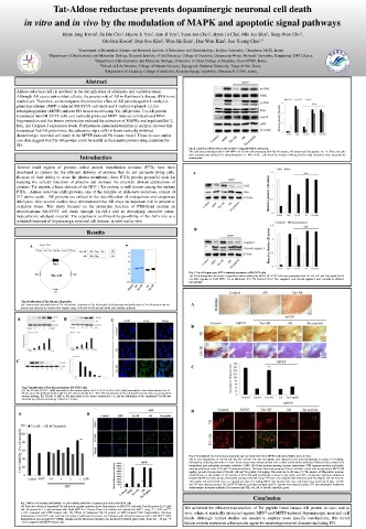
Tat-Aldose reductase prevents dopaminergic neuronal cell death
        in vitro and in vivo by the modulation of MAPK and apoptotic signal pathways

                                     1
                                              1
                   Hyun Jung Kwon , Su Bin Cho , Hyeon Ji Yeo , Eun Ji Yeo , Yeon Joo Choi , Hyun Ju Cha , Min Jea Shin , Sung-Woo Cho ,
                             2
                                                                                1
                                                                                          3
                                                                       1
                                                     1
                                                               1
                                                 5
                                        4
                                                                   2
                                                          1
                               Oh-Shin Kwon , Duk-Soo Kim , Won Sik Eum , Dae Won Kim , Soo Young Choi 1,*
                        1 Department of Biomedical Science and Research Institute of Bioscience and Biotechnology, Hallym University, Chuncheon 24252, Korea.
               2 Department of Biochemistry and Molecular Biology, Research Institute of Oral Sciences, College of Dentistry, Gangneung-Wonju National University, Gangneung 25457, Korea.
                             3 Department of Biochemistry and Molecular Biology, University of Ulsan College of Medicine, Seoul 05505, Korea.
                               4 School of Life Sciences, College of Natural Sciences, Kyungpook National University, Taegu 41566, Korea.
                                5 Department of Anatomy, College of Medicine, Soonchunhyang University, Cheonan-Si 31538, Korea.
                          Abstract
      Aldose reductase (AR) is involved in the detoxification of aldehydes and oxidative stress.
      Although AR exerts anti-oxidant effects, the precise role of AR in Parkinson’s disease (PD) is not
      studied yet. Therefore, we investigated the protective effect of AR protein against 1-methyl-4-
      phenylpyridinium (MPP + )-induced SH-SY5Y cell death and 1-methyl-4-phenyl-1,2,3,6-
      tetrahydropyridine (MPTP)-induced PD mouse model using Tat-AR protein. Tat-AR protein
      transduced into SH-SY5Y cells and markedly protected MPP + -induced cell death and DNA
      fragmentation and this fusion protein also reduced the activation of MAPKs and regulated Bcl-2,
      Bax, and Caspase-3 expression levels. Furthermore, immunohistochemical analysis showed that
      transduced Tat-AR protein into the substantia nigra (SN) in brain markedly inhibited
      dopaminergic neuronal cell death in the MPTP-induced PD mouse model. Those in vitro and in
      vivo data suggest that Tat-AR protein could be useful as therapeutic protein drug candidate for
      PD.
                                                         Fig. 4 Inhibitory effect of Tat-AR on MPP+-induced MAPK activation.
                                                         The cells were stimulated with 5 mM MPP+ for 30 min with pretreated with Tat-AR protein, AR protein and Tat peptide for 1 h. Then, the cells
                         Introduction                    were prepared and analyzed for phosphorylation of JNK, ERK , p38 levels by Western blotting and the band intensities were measured by
                                                         densitometer..
      Several small regions of proteins called protein transduction domains (PTD) have been
      developed as carriers for the efficient delivery of proteins that do not permeate living cells.  A
      Because of their ability to cross the plasma membrane, these PTDs provide powerful tools for
      studying the cellular functions of proteins and increase the potential clinical applications of
      proteins. Tat peptide, a basic domain of the HIV-1 Tat protein, is well known among the various
      PTDs . Aldose reductase (AR) proteins, one of the families of Aldo-keto reductase, consist of
      317 amino acids . AR proteins are critical in the detoxification of endogenous and exogenous
      aldehydes. Also several studies have demonstrated that AR plays an important role in protective
      oxidative stress. This study focused on the protective function of PTD-fused protein on  D
      neuroblastoma SH-SY5Y cell death through Tat-AR’s role in detoxifying oxidative stress-  D
      induced toxic aldehyde material. The experiment confirmed the possibility of Tat-AR’s role as a
      potential treatment of dopaminergic neuronal cell disease, in vitro and in vivo.
                           Results                        B
          A                           B
                                                         Fig. 5 Tat-AR suppresses MPP+-induced apoptosis in SH-SY5Y cells.
                                                         Tat-AR downregulates the levels of apoptotic markers induced by MPP+. SH-SY5Y cells were pretreated with Tat-AR, AR, and Tat peptide for 60
                                                                                   E
                                                         min after exposure to 5mM MPP+ for an additional 18 h. The levels of Bcl-2, Bax, caspase-3, and cleaved caspase-3 were revealed by Western
                                                                                    E
                                                         blot analysis.
       Fig.1 Purification of Tat-AR and AR protein.
       (A) Construction and purification of Tat-AR protein. Overview of Tat-AR protein. (B)Expression and purification of Tat-AR protein and AR  A
       protein were detected by Western blot analysis using 15% SDS-PAGE and anti-rabbit poly histidine antibody.
      A             B             D
                                                         B
      C
                                                         C

       Fig.2 Transduction of Tat-AR proteins into SH-SY5Y cells.
       (A) Tat-AR and AR (0.5–3 μM) was added to the culture medium for 1 h. (B) Tat-AR or AR (3 μM) was added to the culture medium for 15–
       60 min. (C) Cells pretreated with 3 μM Tat-AR were incubated for 1–30 h. The transduction of Tat-AR protein into the cells was analyzed by
       western blotting. (D) Tat-AR (3 μM) or AR was added to the culture medium for 1 h, and the distribution of the transduced Tat-AR was
       observed by confocal microscopy. Scale bar = 20 μm.
                                                         D
      A                    B


                         **

                                                         Fig. 6 Transduced Tat-AR protects dopaminergic neuronal cells from MPTP-induced oxidative stress in vivo.
                                                         (A) In vivo transduction of Tat-AR into the SN. Tat-AR, AR and Tat peptide were injected ip into mice (n=7/group) at a dose of 5.0 mg/kg,
                                                         followed by collecting the brains 6 h later. Brain tissues were immunostained with a rabbit anti-histidine antibody (1:400) and then stained with
                                                         biotinylated goat anti-rabbit secondary antibody (1:200). (B) Brain sections showing tyrosine hydroxylase (TH) immunoreactivity and double
                                                         staining with cresyl violet (CV) and TH immunoreactivity. The brains from each group (n=7) were collected 1 week after ip injection of MPTP (20
                                                         mg/kg), preceded by injection of Tat-AR, AR and Tat peptide (2.0 mg/kg). The scale bar ¼ 100 mm. (C) The number of TH-positive neurons.
                                                         Quantification of the number of DA neurons in 250×250 μm2 (n=7/group) is shown in the graph. ⁎⁎P<0.01, statistically significant difference
                                                         between MPTP and other groups. (D) Double staining of the SN using a TH and a His antibody after treatment with Tat peptide, AR and Tat-AR.
                                           **            Tat peptide, AR and Tat-AR were i.p. injected at a dose of 2 mg/kg, MPTP was injected into mice, and brains were sacrificed 12 later. Tat-AR
                                                         and AR were detected using a His and FITC-labeled secondary antibody and DA neurons were detected using a TH and tetramethyl rhodamine
                                                         isothiocyanate secondary antibody. DA neurons (anti-TH), red; AR or Tat-AR (anti-His), green.
                                                                            Conclusion
       Fig. 3 Effects of transduced Tat-DJ-1 on cell viability and DNA fragmentation in the SH-SY5Y cells.
       (A) Protective effects of transduced Tat-AR protein against oxidative stress. Pretreatment of SH-SY5Y cells with Tat-AR protein (0.5-3 μM)  We achieved the efficient transduction of Tat peptide fused human AR protein in vitro and in
       and AR protein for 1 h and treatment with 5mM MPP + for 13 hours. Then, cell viability was assessed by WST-1 assay. * P < 0.05 and ** P
       < 0.01 compared with MPP + -treated cells. (B) Effects of transduced Tat-AR protein on MPP + -induced DNA fragmentation. One-hour  vivo, where it markedly protected against MPP + -and MPTP-induced dopaminergic neuronal cell
       pretreatment of SH-SY5Y cells with Tat-AR protein (3 μM) and AR protein was followed with 16 hours treatment with 5 mM MPP + . DNA
       fragmentation was measured by TUNEL staining and the fluorescent intensity was measured by ELISA plate reader. Scale bar = 50 μm. ** P  death. Although further studies are needed to explore more specific mechanisms, this novel
       < 0.01 compared with MPP + -treated cells.       fusion protein represents a therapeutic agent for neurodegenerative diseases including PD.