
Page 5 - Q. Neuroscience
P. 5
MiR-30 and miR-153 alleviate LPS induced inflammation by targeting NeuroD1 in microglial cells.
2*
1
1
Hye-Rim Choi , Ji Sun Ha , Sung-Woo Cho , Seung-Ju Yang 1*
1 Department of Biomedical Laboratory Science, Konyang University, Daejeon, 35365, Korea
2 Department of Biochemistry and Molecular Biology, University of Ulsan College of Medicine, Seoul, Korea
BACKGROUND AIM
Neurogenic differentiation 1 (NeuroD1) is a basic helix-loop-helix (bHLH) In the present study, we aimed to analyze the immunoregulatory
transcription factor that plays an important role during neuronal differentiation, mechanisms of NeuroD1 in BV-2 cells induced by LPS. Therefore, we
maturation and survival. It is reported that NeuroD1 is associated with confirmed anti-inflammatory effects by inhibiting NeuroD1, which is
inflammatory response in LPS-induced BV-2 cells. MicroRNA (miRNA) is increased in LPS-induced BV-2 cells. Moreover, NeuroD1 targeting
endogenous small non-coding RNAs consisting of 22 nucleotides that miRNAs are constructed and assessed its effectiveness. After then, we
effectively regulates gene expression at the translation level. It is related to a investigate the anti-inflammatory effects of miRNAs against LPS induced
variety of central nervous system diseases and microglia differentiation. inflammation responses and NLRP3 inflammasome activation in BV-2 cells.
METHODS
1) Cell culture
: BV-2 cells were maintained in DMEM with penicillin (100 U/mL), streptomycin (100 μg/mL), and 10% FBS in a humidified incubator at 37°C in 5% CO 2 .
2) Plasmids and transfection
: Cells were transfected with NeuroD1 shRNA, scramble shRNA and microRNA using lentiviral packaging vector. Cells were incubated for 24 h before other experiments.
3) Western blotting
: BV-2 cells were lysed using RIPA buffer. Proteins were separated on 10% - 12% SDS gels by electrophoresis. The proteins were transferred onto a nitrocellulose membrane,
and then incubated overnight at 4℃ with the primary antibody, followed by incubation for 1h at room temperature with secondary antibody.
4) Real-time PCR
: RNA were extracted using Trizol reagent according to manufacturer’s instructions. After then synthesize the cDNA using RT-PCR and RT-qPCR was performed.
RESULTS
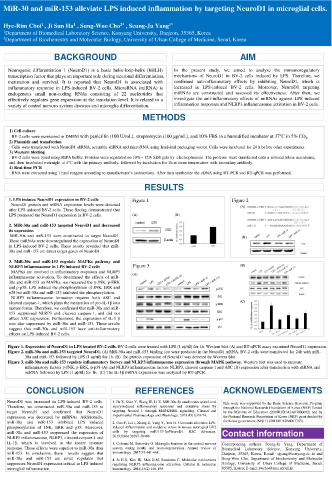
1. LPS induces NeuroD1 expression in BV-2 cells Figure 1 Figure 2
NeuroD1 protein and mRNA expression levels were detected (A)
after LPS induced BV-2 cells. These finding demonstrated that
LPS promoted the NeuroD1 expression in BV-2 cells. (A) (B)
2. MiR-30a and miR-153 targeted NeuroD1 and decreased
its expression
MiR-30a and miR-153 were constructed to target NeuroD1. (B)
These miRNAs were downregulated the expression of NeuroD1
in LPS-induced BV-2 cells. These results revealed that miR-
30a and miR-153 are direct target genes of NeuroD1.
3. MiR-30a and miR-153 regulate MAPKs pathway and
NLRP3 inflammasome in LPS induced BV-2 cells Figure 3 (B)
MAPKs are involved in inflammatory responses and NLRP3
inflammasome activation. To determined the effects of miR- (A)
30a and miR-153 on MAPKs, we measured the p-JNK, p-ERK
and p-p38. LPS induced the phosphorylation of JNK, ERK and
p38 but miR-30a and miR-153 inhibited the phosphorylation.
NLRP3 inflammasome formation requires both ASC and
cleaved caspase-1, which plays the maturation of pro-IL-1β into (C)
mature forms. Therefore, we confirmed that miR-30a and miR-
153 suppressed NLRP3 and cleaved caspase-1, and did not
affect ASC expression. Furthermore, the expression of IL-1 β
was also suppressed by miR-30a and miR-153. These results
suggest that miR-30a and miR-153 have anti-inflammatory
effects on LPS-induced BV-2 cells.
Figure 1. Expression of NeuroD1 in LPS treated BV-2 cells. BV-2 cells were treated with LPS (1 ㎍/㎖) for 1h. Western blot (A) and RT-qPCR assay examined NeuroD1 expression.
Figure 2. miR-30a and miR-153 targeted NeuroD1. (A) MiR-30a and miR-153 binding site were predicted in the NeuroD1 mRNA. BV-2 cells were transfected for 24h with miR-
30a and miR-153 followed by LPS (1 ㎍/㎖) for 1h. (B) The protein expression of NeuroD1 was detected by Western blot.
Figure 3. miR-30a and miR-153 regulate inflammatory factors and NLRP3 inflammasome expression through MAPK pathway. Western blot was used to measure
inflammatory factors p-JNK, p-ERK, p-p38 (A) and NLRP3 inflammasome factors NLRP3, cleaved caspase-1 and ASC (B) expression after transfection with shRNA and
miRNA followed by LPS (1 ㎍/㎖) for 1h. (C) The IL-1β mRNA expression was analyzed by RT-qPCR.
CONCLUSION REFERENCES ACKNOWLEDGEMENTS
NeuroD1 was increased in LPS-induced BV-2 cells. 1. Fu X, Shen Y, Wang W, Li X. MiR‐30a‐5p ameliorates spinal cord This work was supported by the Basic Science Research Program
Therefore, we constructed miR-30a and miR-153 to injury‐induced inflammatory responses and oxidative stress by through the National Research Foundation of Korea (NRF) funded
target NeuroD1 and confirmed that NeuroD1 targeting Neurod 1 through MAPK/ERK signalling. Clinical and by the Ministry of Education (2018R1D1A3A03000692) and by
expression was decreased by miRNAs. Additionally, Experimental Pharmacology and Physiology. 2018;45(1):68-74. the National Research Foundation of Korea (NRF) grant funded by
miR-30a and miR-153 inhibited LPS induced 2. Gao F, Lei J, Zhang Z, Yang Y, You H. Curcumin alleviates LPS- the Korea government (MSIT) (2018R1A2B6001743).
phosphorylation of JNK, ERK and p38. Moreover, induced inflammation and oxidative stress in mouse microglial BV2
miR-30a and miR-153 suppressed the expression of cells by targeting miR-137-3p/NeuroD1. RSC Advances. Contact information
NLRP3 inflammasome, NLRP3, cleaved caspase-1 and 2019;9(66):38397-38406.
IL-1β, which is involved in the innate immune 3. Colonna M, Butovsky O. Microglia function in the central nervous Corresponding authors. Seung-Ju Yang, Department of
response. These effects were superior to miR-30a than system during health and neurodegeneration. Annual review of Biomedical Laboratory Science, Konyang University,
miR-153. In conclusion, these results suggest that immunology. 2017;35:441-468. Daejeon, 35365, Korea; E-mail: sjyang@konyang.ac..kr and
miR-30a and miR-153 are novel regulator that 4. Jo E-K, Kim JK, Shin D-M, Sasakawa C. Molecular mechanisms Sung-Woo Cho, Department of biochemistry and Molecular
suppresses NeuroD1 expression critical in LPS induced regulating NLRP3 inflammasome activation. Cellular & molecular Biology, University of Ulsan College of Medicine, Seoul,
microglial inflammation. immunology. 2016;13(2):148-159. 05505, Korea; E-mail: swcho@amc.seoul.kr