
Page 13 - Q. Neuroscience
P. 13
MicroRNAs in serum-derived neuronal exosomes as biomarkers of
acute severe stress response
Minkyoung Sung¹ , Soo-Eun Sung¹ , Kyung-Ku Kang¹, Joo-Hee Choi¹, Si-Joon Lee¹, Kil-Soo Kim¹, Min-Soo Seo¹ *
,#
,
,#
Laboratory animal center, Daegu Gyeongbuk Medical Innovation Foundation, Daegu, Korea
Abstract
Stress is the physical and psychological tension that individual feels when faced with a situation that is difficult to adapt. Previous studies have shown that stress alters the
expression of stress hormones and then causes brain neuroinflammation. We further analyzed the miRNAs in serum derived neuronal exosome to confirm that miRNAs
with different expression levels after exposure to acute stress can be used as stress biomarkers. First, each stress protocol was treated to make stress animal models
according to different stress severity. Next, we analyzed stress hormones such as corticosterone, cortisol and neuron-associated inflammation marker such as BDNF, COX2,
GFAP and TNF-α according to stress severity and time. Because control and severe group were showed significant differences among them, neuronal exosomes were
isolated from serum of control and severe group. Following exosomes isolation, NGS was performed to measure exosomal miRNA of severe group against exosomal miRNA
of control group. As a result, 13 upregulated miRNAs and 11 downregulated miRNAs were analyzed. Many studies have shown that several miRNAs among 24 miRNAs
regulate neuron and depression-associated factors. Thus, these miRNAs in serum derived neuronal exosomes may be used as biomarkers of stress response.
Introduction A B CD9 CD63
98.96% 78.36%
Stress is defined as tension that an individual accepts when placed in an
environment that is difficult to adapt to or threatened to maintain homeostasis.
Glucocorticoid hormones such as cortisol, corticosterone have used as biomarkers
of psychological stress, but the previous studies indicated that the stress response
of HPA axis is influenced by many factors. So many studies are underway to find C D Size distribution by intensity
biomarkers that can objectively judge stress. Stress is known to affect the brain tEV
and alter gene expression of neuroinflammatory markers in the hippocampus, CD9
which is particularly sensitive to stress. Also, exosomes and exosomal miRNAs CD81
have studied as diagnostic biomarkers of many diseases. Based on these data, this TSG101
study aims to identify neuronal exosomal miRNAs that have potential as stress
biomarkers by confirming miRNAs whose expression levels change with stress. Figure 3. Characterization of total exosomes isolated from serum. (A) TEM image
showing exosome morphology and size. (B) FACS data confirming expression of
Materials and Methods exosome markers. (C) Western blot analysis for confirm of exosome markers. (D)
DLS analysis of exosomes to confirm size distribution.
Severe stress was induced by Figure 4. Characterization of neuronal exosomes
electronic foot shock.
Exosomes were isolated from isolated from serum. Western blot image shows
enrichment of neuronal exosome-associated marker.
serum using Exo-Quick solution.
Neuronal exosomes were isolated
by referring to Mustapic, Maja, et
al.
Analysis of microRNAs was performed by next-generation sequencing (NGS).
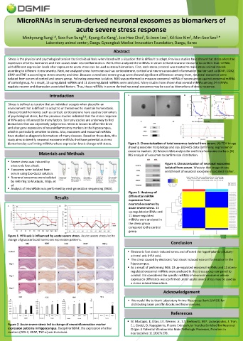
Figure 5. Heatmap of
Results differential miRNA
expression from
neuronal exosomes by
acute severe stress. 13
up-regulated miRNAs and
11 down-regulated
miRNAs were analyzed in
the stress group
compared to the control
group.
Figure 1. HPA axis is influenced by acute severe stress. Acute severe stress led to
change of glucocorticoid hormones expression patterns.
Conclusion
Electronic foot shock-induced stress and affected the hypothalamic-pituitary-
adrenal axis (HPA axis).
The stress caused by electronic foot shock induced neuroinflammation in the
hippocampus.
As a result of performing NGS, 13 up-regulated exosomal miRNAs and 11 down-
regulated exosomal miRNAs were analyzed in the stress group compared to
control. It is considered the specific miRNAs of neuronal exosomes whose
expression difference was confirmed under acute severe stress may be used as
a stress-related biomarkers.
Acknowledgement
We would like to thank Laboratory Animal Resources Bank (LAREB) for
distributing brain paraffin blocks and frozen serums.
References
M. Mustapic, E. Eitan, J.K. Werner, Jr., S.T. Berkowitz, M.P. Lazaropoulos, J. Tran,
Figure 2. Acute severe stress led to change of neuroinflammation marker E.J. Goetzl, D. Kapogiannis, Plasma Extracellular Vesicles Enriched for Neuronal
expression patterns in hippocampus. Except for BDNF, the expression of other Origin: A Potential Window into Brain Pathologic Processes, Frontiers in
markers (COX-2, GFAP, TNF-α) was increased. neuroscience 11 (2017) 278.