
Page 17 - Q. Neuroscience
P. 17
Reciprocal connections between mediodorsal thalamus and prefrontal cortex
Yelin Lee, Hyeonyeong Jeong, Eunji Cheong*
Department of Biotechnology, College of Life Science and Biotechnology, Yonsei University, Seoul 120-752, Korea
BACKGROUND
Prefrontal cortex (PFC)–mediodorsal thalamus (MD) interactions are critical for cognition. In addition, executive functions, such as working memory and cognitive control,
majorly depend on PFC integrity. Most of the current knowledge about the thalamocortical (TC) projection organization has been obtained by relying on neuronal circuit-
tracing approaches; however, the underlying circuits are unclear, and the results of previous studies are inconsistent. Therefore, to investigate the connections between
the MD and various PFC regions, this study focused on specific MD–PFC connections using anterograde and retrograde tools. Anterograde and retrograde tools were
injected into the MD of mice, and different MD and PFC subdivisions were examined. In addition, the quantitative and qualitative features of the MD–medial prefrontal
cortex (mPFC) pathway were analyze using direct comparisons of labeling. Different MD subdivisions characterized differential TC connectivity. The MD received
excitatory inputs from the motor region (primary and secondary motor cortex [M1 and M2]), mPFC (anterior cingulate area, prelimbic and infralimbic areas), orbitofrontal
cortex, and agranular insular cortex and inhibitory inputs from the reticular nucleus of the thalamus and the zona incerta. The results provided insight into a new
corticothalamic circuit and the consciousness-related function of the MD, which will help understand the TC connectivity of this region and determine its functional role.
AIM
MD plays a role in memory and other cognitive tasks. Anatomical tracing studies have confirmed at least three different MD subdivisions: medial MD (MDm), central MD
(MDc), and lateral MD (MDl). Each is differentially interconnected to the PFC. The most anterior part of frontal lobes is the part of the cortex innervated primarily by
afferents from the mediodorsal nucleus of the thalamus. Interest in understanding the functional contribution of thalamic inputs to cortical functions and of different
thalamic afferents has significantly increased as it may provide insight into the relative specialization of PFC subdivisions. This study focused on specific connections
from the MD to the PFC through direct comparisons of labeling produced by anterograde and retrograde tracing injections in different subdivisions of the PFC in mice.
The differential TC connectivity characterizes different clusters of cells in the MD, and MD projections to the PFC are related to different MD neuronal populations. The
data collected will reveal the neuronal architecture of MD projections to the PFC and provide a better understanding of the quantitative and qualitative features of MD–
PFC pathways. In addition, a clearer anatomical and functional understanding of thalamic-frontal circuitry will clarify how its alteration might contribute to cognitive
dysfunction in psychiatric conditions.
METHODS RESULTS
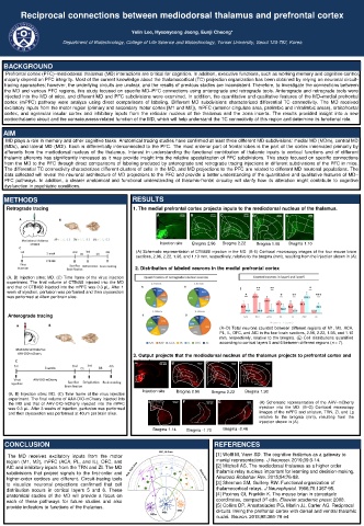
Retrograde tracing 1. The medial prefrontal cortex projects inputs to the mediodorsal nucleus of the thalamus.
A B C D E
l c
m
Injection site Bregma 2.96 Bregma 2.22 Bregma 1.98 Bregma 1.10
(A) Schematic representation of CTB488 injection in the MD. (B-E) Confocal microscopy images of the four mouse brain
sections, 2.96, 2.22, 1.98, and 1.10 mm, respectively, relative to the bregma (mm), resulting from the injection shown in (A).
2. Distribution of labeled neurons in the medial prefrontal cortex
(A, B) Injection sites: MD. (C) Time frame of the virus injection
experiment. The final volume of CTB488 injected into the MD
and that of CTB488 injected into the mPFC was 0.3 μL. After 1
week of injection, perfusion was performed and then cryosection
was performed at 40um per brain slice.
Anterograde tracing
(A–D) Total neurons counted between different regions of M1, M2, ACA,
PL, IL, OFC, and AIC in the four brain sections, 2.96, 2.22, 1.98, and 1.10
mm, respectively, relative to the bregma. (E) Cell distributions quantified
according to cortical layers 5 and 6 between different regions (n = 7).
3. Output projects that the mediodorsal nucleus of the thalamus projects to prefrontal cortex and
A subcortex. B C D
Injection site Bregma 2.96 Bregma 2.22 Bregma 1.98
(A, B) Injection sites: MD. (C) Time frame of the virus injection
experiment. The final volume of AAV-DIO-mCherry injected into
the MD and that of AAV-DIO-mCherry injected into the mPFC E F G (A) Schematic representation of the AAV–mCherry
was 0.3 μL. After 3 weeks of injection, perfusion was performed injection into the MD. (B–G) Confocal microscopy
and then cryosection was performed at 40um per brain slice. images of the mPFC and striatum, TRN, ZI, and Lg
relative to the bregma (mm), resulting from the
injection shown in (A).
Bregma 1.14 Bregma -1.70 Bregma -2.46
CONCLUSION REFERENCES
The MD receives excitatory inputs from the motor [1] Wolff M, Vann SD. The cognitive thalamus as a gateway to
region (M1, M2), mPFC (ACA, PL, and IL), OFC, and mental representations. J Neurosci. 2019;39:3-14.
AIC and inhibitory inputs from the TRN and ZI. The MD [2] Mitchell AS. The mediodorsal thalamus as a higher order
subdivisions that project signals to the first-order and thalamic relay nucleus important for learning and decision-making.
higher-order cortices are different. Circuit-tracing tools Neurosci Biobehav Rev. 2015;54:76-88.
to visualize neuronal projections confirmed that cell [3] Sherman SM, Guillery RW. Functional organization of
distribution occurs in cortical layers 5 and 6. These thalamocortical relays. J Neurophysiol. 1996;76:1367-95.
anatomical studies of the MD will provide a focus on [4] Paxinos Gf, Franklin K. The mouse brian in stereotaxic
rd
each of these pathways for future studies and also coordinates, compact 3 edn. Elsevier academic press: 2008.
provide indicators to functions of the thalamus. [5] Collins DP, Anastasiades PG, Marlin JJ, Carter AG. Reciprocal
circuits linking the prefrontal cortex with dorsal and ventral thalamic
nuclei. Neuron. 2018;98:366-79 e4