
Page 5 - M. Immunology
P. 5
The function of microRNA-155 and Chronic Chorioamnionitis in Swan 71,
human trophoblasts.
Hee-Na Jeong1,#, So-Yul Eom1,#, Sun-Shim Choi1, Hae-Ryeong Lim1,** and Deug-Chan Lee1,*
1Department of Medical biotechnology, College of Biomedical Science, Kangwon National University, Republic of Korea
BACKGROUND AIM
Chronic choroiditis (CCA) is an infectious pregnancy disease caused by maternal immune cells MicroRNAs are non-coding RNA, single-stranded RNA, consisting of 22 nucleotides that target mRNA by binding to complementary
infiltrate into the amnion surrounding the fetus and the chorion that invade the endometrium. The sequences to induce RNA silencing. Chronic choroiditis (CCA) is a pregnancy disease caused by maternal immune cells infiltrating
prevalence of CCA is 34% of all preterm, 39% of all preterm due to prelabor rupture of the membranes into the amnion surrounding the fetus and the chorion that invade the endometrium. The prevalence of CCA is 34% of all preterm,
(PROM) and 60% of fetal mortality but has no significance. Recently, the study of microRNAs 39% of all preterm due to prelabor rupture of the membranes (PROM) and contributes to fetal death. In this study, we confirmed the
regulated with pregnancy has increased and the molecular mechanisms by which microRNAs regulate fetal membrane of CCA, Acute Chorioamnionitis (ACA) and No-inflammation (normal) and found that miR-155 was overexpressed in
pregnancy or its associated disease were being reported. However, there is a lack of research the fetal membranes of CCA. Also differentially expressed genes (DEGs) by elevated miR-155 in trophoblasts that constitute the fetal
regarding microRNA and CCA. Karyopherin α1 (KPNA1) is subunit of nuclear transport complex, membrane were investigated using microarray analysis. Based on the results, we focus on the function of KPNA1 which is known to
especially contributes transport phosphorylation of Signal transducer and activator of transcriptions function as a nuclear transporter of the phosphorylated-signal transducer and activator of transcription 3 (p-STAT3) homodimer into
(STATs). KPNA1 mRNA is highly expressed in the placenta, but its function is unknown. STAT3 the nucleus. Signal transducer and activator of transcription 3 (STAT3) are intracellular transcription factors involved in cell migration
signaling plays a key role in many cellular processes such as cell growth and apoptosis and controls in and cell apoptosis and have been reported that it was suppressed in inflammation of a fetus caused by CCA. We confirmed that miR-
the development of embryonic. Also, It have been reported that it was suppressed in inflammation of a 155 directly targets KPNA1, result in suppression of KPNA1 protein. Knock-down of KPNA1 inhibits the translocation of
fetus caused by CCA. In this study, we investigate expression of miRNAs in tissue of pregnancy- phosphorylated STAT3 into the nucleus. In conclusion, we demonstrated that miR-155 targets KPNA1 and thereby inhibits STAT3
related diseases and profiled miR-155 in human trophoblasts. signaling in human trophoblast cell, Swan 71.
METHODS
<Trophoblasts transfection and RNA isolation> Swan 71 cells, human trophoblasts were cultured in Dulbecco’s modified Eagle’s medium supplemented with 10% heat inactivated fetal bovine serum, 1% penicillin streptomycin. The Cell is incubated in a humidified atmosphere
containing 5% CO₂ at 37℃. To over-express miR-155 for microarray analysis or to induce KPNA1 knock-down, 1.5 × 10⁶ Swan 71 cells were transfected with 25 pmole of hsa-miR-155-5p or 25 pmole of KPNA1 siRNA using the Neon transfection system (1,050 mV, 30 ms and
two pulses) and were harvested after 12, 24 and 48 hours for immunoblotting, 48 hours for microarray and qRT-PCR validation. The RNA Total RNA for qRT-PCR was isolated using miRNeasy mini kit. <Microarray and data analysis> Microarray analysis was performed with the
standard protocol by DNA Link incorporation, an Affymetrix authorized service provider (Seoul, Republic of Korea). For the gene expression profiles of miR-155 transfected samples, hybridization and staining were performed with GeneChip Human Gene 2.0 ST Arrays
(Affymetrix). After acquiring the array images by the Affymetrix GeneChip Scanner 30007G, the intensity of acquired images were analyzed using Affymetrix GCOS 1.4 software. The normalization used a robust multi-array average (RMA) algorithm, Affymetrix Expression
Console 1.3.1v and then these data were transformed into 1og2 scale. Differentially expressed genes (DEGs) were defined that the genes significantly (p < 0.05) expressed more than 1.5-fold change in miR-155 transfected samples, compared to negative control miRNA
transfected samples. And hierarchical clustering analysis was conducted by using R package to confirm whether the previous experiments were correctly processed. To further analyze DEGs, gene-biological pathway analysis was performed and visualized using Cytoscape.
Quantitative real-time reverse transcription polymerase chain reaction> he same RNA used for the microarray analysis was measured using the EpochTM microplatereader and 200 ng of RNA was reverse-transcribed to cDNA using ReverTra Ace qPCR RT Master Mix. qRT-
PCR was conducted to determine comparative mRNA expression using THUNDERBIRD SYBR qPCR Mix. The primer pairs of miR-155 target genes were synthesized by Cosmo (Seoul, Republic of Korea), showing Table 1. For normalization, a house keeping gene HPRT was
used, which is not affected by miR-155. qRT-PCR was conducted using a StepOne Plus Real-Time PCR system (Applied Biosystems). The fold change was derived using the ΔΔCt method : △Ct = Ct target gene – Ct internal reference (RPLP0) and △△Ct = △Ct sample – △Ct
control. <Immunoblotting> Total proteins were isolated from harvested cell pellets using RIPA buffer containing protease and phosphatase inhibitor and nuclear and cytoplasmic proteins were isolated from NE-PER Nucelar and Cytoplasmic extraction Reagents. 30 ㎍ of total
cellular proteins and 10 ㎍ of nuclear and cytoplasmic proteins were subjected to 12% SDS-PAGE gel electrophoresis and electrotransferred onto PVDF membrane. The membranes were blocked with 5% skim milk in TBS-T for an hour and then were incubated overnight at 4℃
with a specific primary antibodies. After washing three-times with TBS-T, the membranes were incubated with hourseradish peroxidase (HRP)-conjugated secondary antibodies in 1% skim milk for 2 hours. After washing three-times with TBS-T, the membranes were detected
using WesternBright Peroxide chemiluminescent detection reagent. <Dual-luciferase assay> To confirm whether miR-155 directly binds KPNA1, the KPNA1 3’ untranslated region (UTR) DNA fragment was amplified from Swan 71 cell total RNA using a designed primer by RT-
PCR and cloned into T-Vector pMD20. Co-transfection with 25pmole hsa-miR-155-5p or mirVana miRNA, pMIR-KPNA1 3’ UTR and pRL-SV40 plasmid in Swan 71 cells were performed using the Neon transfection system, The Dual-Luciferase Reporter Assay System was used
to measure the firefly and renilla luciferase activity following the manufacturer’s protocol. Renilla luciferase activity was defined as an internal control. <Statistical analysis> Each experiment was conducted independently at least three times. All the result in this study have been
expressed as the mean ± standard deviation (SD). Statistical differences between control groups and sample groups were analyzed by Student’s t-test using SPSS. Differences with p-value < 0.05 were considered statistically significant.
RESULTS
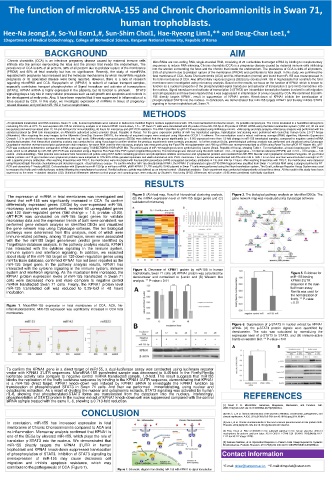
The expression of miRNA in fetal membranes was investigated and Figure 2. (A) Heat map, Result of hierarchical clustering analysis, Figure 3. The biological pathway analysis on identified DEGs. The
gene network map was visualized using Cytoscape software
(B) the mRNA expression level of miR-155 target genes and (C)
found that miR-155 was significantly increased in CCA. To confirm validation of microarray
differentially expressed genes (DEGs) by over-expressed miR-155,
microarray analysis was performed, revealed 90 up-regulated genes
and 120 down-regulated genes (fold change > 1.5, p-value <0.05).
qRT-PCR was conducted on miR-155 target genes for validate
microarray data and the expression trends of both were consistent. we
performed gene network analysis on identified DEGs and visualized
the gene network map using Cytoscape software. The ten biological
pathways were determined from this analysis, most of which were
immune-related pathway. among 10 pathways, seven were associated
with the five miR155 target gene/seven predict gene identified by
TargetScan database analysis. In the pathway analysis results, KPNA1
has interacted with the cytokine signaling in the immune system,
immune system and interferon signaling. In addition, we searched
about study of the miR-155 target on 120 down-regulation genes using
miRTarBase database, confirmed KPNA1 has not been reported as the
miR-155 target gene. In the pathway analysis results, KPNA1 has
interacted with the cytokine signaling in the immune system, immune Figure 4. Decrease of KPNA1 protein by miR-155 in human
system and interferon signaling. As the incubation time increased, the trophoblasts, Swan 71 cells. (A) KPNA1 protein was calculated by Figure 5. Evidence for
KPNA1 protein expression levels of miR-155 transfected in Swan 71 densitometer and normalized to β-actin and (B) Western blot miR-155 binding
cells were decreased more and more compare to negative control analysis. ** P-value < 0.01 KPNA1 3’UTR
miRNA transfected Swan 71 cells. Finally, the KPNA1 protein level sequence in the dual-
miR-155 transfected cell was reduced to 0.29-fold in 48 hours luciferase assay.
incubation. Renilla was used for
the normalization of
firefly. ** P-value
< 0.01
Figure 1. MicroRNA-155 expression in fetal membranes of CCA, ACA, No-
inflammation(control). MiR-155 expression was significantly increased in CCA fetal
membranes.
Figure 6. Suppression of p-STAT3 in nuclear extract by KPNA1
siRNA. (A) the p-STAT3 protein signals were quantified by
densitometer. The ratio was calculated by normalizing the
expression level of p-STAT3 to STAT3. and (B) immuno-active
bands on western blot. ** P-value < 0.01
To confirm the KPNA1 gene is a direct target of miR155, a dual-luciferase assay was conducted using luciferase reporter
vector with KPNA1 3’UTR sequences. MicroRNA-155 transfected sample was decreased to 0.45-fold in the Firefly/Renilla
luciferase activity ratio compare to negative control miRNA transfected sample, 1.0-fold. This result suggests that miR155
blocks the replication of the firefly luciferase sequence by binding to the KPNA1 3′UTR sequence, demonstrating that KPNA1
is a miR-155 direct target. KPNA1 knock-down was induced by KPNA1 siRNA to investigate the KPNA1 function as
translocation of phosphorylated STAT3 in Swan 71 cells. And then we performed immunoblotting, using nuclear and
cytoplasmic extraction. As a result of dividing the nuclear and cytoplasmic extracts, STAT3 signaling was activated by human
rIL-6, showing that phosphorylated-STAT3 forms are translocated from the cytoplasm into the nucleus. Interestingly, REFERENCES
phosphorylation of STAT3 protein in the nuclear extract of KPNA1 knock-down cell was suppressed compared with the control
siRNA sample treated with the same IL-6, showing a 0.713-fold reduction.
Biogenesis,
[1] 2004;116(2):281-297. doi:10.1016/S0092-8674(04)00045-5. Mechanism, and Function. Cell.
David P. B. MicroRNAs:
Genomics,
CONCLUSION [2] Kim C. J, et al. Chronic inflammation of the placenta: definition, classification, pathogenesis, and
clinical significance. AJOG. 2015;213(4):S53-S69.doi:10.1016/j.ajog.2015.08.041.
In conclusion, miR-155 has increased expression in fetal [3] Lee J, et al. Chronic chorioamnionitis is the most common placental lesion in late preterm birth.
Placenta. 2013;34(8):681-689. doi:10.1016/j.placenta.2013.04.014.
membranes of Chronic Chorioamnionitis compared to ACA and
no-inflammation. Microarray analysis confirmed that KPNA1 is [4] Zhou Xin,et al. Role of SOCS3 in the Jak/stat3 pathway in the human placenta: different
mechanisms for preterm and term labor. ACTA OBSTET GYNECOL SCAND. 2015;94(10):1112-
one of the DEGs by elevated miR-155, which plays the role of 1117.doi:10.1111/aogs.12708.
translation p-STAT3 into the nucleus. We demonstrated that [5] Seehase Matthias, et al. Myocardial Response in Preterm Fetal Sheep Exposed to Systemic
miR-155 directly targets the KPNA1 3’UTR in human Endotoxinaemia. Pediatric Research. 2011;70(3):242-246. doi:10.1203/PDR.0b013e318225fbcb.
trophoblast and KPNA1 knock-down suppressed translocation
of phosphorylation of STAT3. Inhibition of STAT3 signaling by Contact information
overexpression of miR-155 may cause decreases cell
migration and inhibits apoptosis resistance, which may
contribute to the pathogenesis of CCA (Figure 7). *E-mail: dclee@kangwon.ac.kr, **E-mail: dimgofud@naver.com
Figure 7. Schematic diagram from binding miR-155 with KPNA1 to signal transduction