
Page 3 - M. Immunology
P. 3
Identification of the in vivo role of Tetraspanin 7
in osteoclastic bone resorption
Minhee Kim* and Soo Young Lee
Department of Life Science, Ewha Womans University, Seoul 03760, Republic of Korea
BACKGROUND AIM
Bone is a highly dynamic organ, which is constantly being remodeled Our group previously identified that Tspan7 is highly upregulated by
1
throughout life by two major types of cells, osteoblasts and osteoclasts . RANKL stimulation in the late stage of osteoclastogenesis and is
Osteoblasts enhance new bone formation and osteoclasts resorb bone ma necessary for mature osteoclast function (unpublished, L. Jing et al).
2
-trix . After bone is resorbed by osteoclasts, it is subsequently followed by Furthermore, Tspan7 links RANK and αvβ3 integrin complex through
3
the replacement of osteoblasts . The imbalance between osteoblasts and its specialized structure, TEM. Kwon et al. also described that Tspan7
9
osteoclasts results in skeletal diseases such as osteoporosis, osteoarthritis, modulates cytoskeleton rearrangement and bone resorption in vitro .
4, 5
-/-
and cancer-associated bone diseases . Recently, how tetraspanins affect In this study, we generated Tspan7-knockout (Tspan7 ) mice using
the development and maintenance of the skeletal system has been a focus CRISPR/Cas9 system to investigate the role of Tspan7 in
on studies. For example, CD9 and Tspan5 positively regulate osteoclast for osteoclastogenesis in vivo.
6, 7
mation during osteoclastogenesis in vitro . A more recent study showed
that CD82 is essential for osteoclast fusion and osteoclast function through
8
cytoskeleton regulation in vivo .
METHODS
-/-
+/
For this study, Tspan7 mice were generated using CRISPR/Cas9 system. Mice were produced by crossing Tspan7 +/− females with Tspan7 Y males or
−/
Tspan7 Y males. All mice were genotyped 10 days after birth using toe clipping. For osteoclast formation, isolated BMMs were grown with M-CSF
(30ng/ml) and RANKL (100ng/ml) for 4-5 days, and mature osteoclasts were stained with TRAP. To analyze osteoclast function, pre-osteoclasts were
seeded onto dentin slices with the culture medium containing M-CSF (30ng/ml) and RANKL (100ng/ml) for 2 days. Adherent cells on dentin slices
were removed and stained with hematoxylin. For actin ring formation, mature osteoclasts grown on poly-L-lysine-coated glass coverslips and dentin
-/-
slices stained with phalloidin. To establish LPS-induced calvarial bone destruction model, 5-6 weeks old male WT and Tspan7 mice were randomly
divided into two groups (n = 4-5/group). Either PBS or LPS was injected into the subcutaneous tissue of the calvaria twice at 2 days interval. Five
days after the first injection, the mice were sacrificed. Calvarial bone sections were processed for TRAP staining, H&E staining and
-/-
immunohistochemistry. For OVX-induced postmenopausal osteoporosis model, 10-11 weeks old female WT and Tspan7 mice were performed
bilateral ovariectomy (OVX) or sham-operation. Seven weeks after operation, the mice were sacrificed. Then, bone histomorphometry were
conducted as previously described.
RESULTS
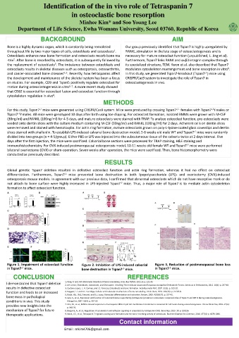
Global genetic Tspan7 deletion resulted in defective osteoclast function and actin ring formation, whereas it had no effect on osteoclast
differentiation. Furthermore, Tspan7 -/- mice prevented bone destruction in both lipopolysaccharide (LPS)- and ovariectomy (OVX)-induced
osteoporosis mouse models. In agreement with our previous data, I confirmed that abnormal osteoclasts which do not have resorptive mark or do
-/-
not attach to bone surface were highly increased in LPS-injected Tspan7 mice. Thus, a major role of Tspan7 is to mediate actin cytoskeleton
formation to affect osteoclast function.
Figure 1. Impairment of osteoclast function Figure 2. Inhibition of LPS-induced calvarial Figure 3. Reduction of postmenopausal bone loss
-/-
-/-
in Tspan7 mice. bone destruction in Tspan7 mice. in Tspan7 mice.
-/-
CONCLUSION REFERENCES
1. Feng, X. and J.M. McDonald, Disorders of bone remodeling. Annu Rev Pathol, 2011. 6: p. 121-45.
I demonstrated that Tspan7 deletion
2. UH Lerner, Osteoblasts, Osteoclasts, and Osteocytes: Unveiling Their Intimate-Associated Responses to Applied Orthodontic Forces. Seminars in Orthodontics, 2012. 18(4): p. 237-48.
results in defective osteoclast 3. Caetano-Lopes, J., H. Canhao, and J.E. Fonseca, Osteoblasts and bone formation. Acta Reumatol Port, 2007. 32(2): p. 103-10.
function and leads to an increased 4. Raggatt, L.J. and N.C. Partridge, Cellular and molecular mechanisms of bone remodeling. J Biol Chem, 2010. 285(33): p. 25103-8.
5. Boyle, W.J., W.S. Simonet, and D.L. Lacey, Osteoclast differentiation and activation. Nature, 2003. 423(6937): p. 337-42.
bone mass in pathological 6. Iwai, K., et al., Expression and function of transmembrane-4 superfamily (tetraspanin) proteins in osteoclasts: reciprocal roles of Tspan-5 and NET-6 during osteoclastogenesis.
conditions in vivo. This study Allergol Int, 2007. 56(4): p. 457-63.
provides new insights into the 7. Ishii, M., et al., RANKL-induced expression of tetraspanin CD9 in lipid raft membrane microdomain is essential for cell fusion during osteoclastogenesis. J Bone Miner Res, 2006. 21(6):
p. 965-76.
mechanism of Tspan7 for future 8. Bergsma, A., et al., Regulation of cytoskeleton and adhesion signaling in osteoclasts by tetraspanin CD82. Bone Rep, 2019. 10: p. 100196.
therapeutic applications. 9. Kwon, J.O., et al., Tetraspanin 7 regulates sealing zone formation and the bone-resorbing activity of osteoclasts. Biochem Biophys Res Commun, 2016. 477(4): p. 1078-1084.
Contact information
Email : mkim4706@gmail.com