
Page 29 - M. Immunology
P. 29
Sirtuin 3 activation enhances autophagy and antimicrobial responses and
ameliorates mitochondrial damage and oxidative stress during Mtb infection
Hyun-Woo Suh 1,2,3 , Tae Sung Kim 1,2,3 , Jin Kyung Kim 1,2,3 , Jin Ho Choe 1,2,3 , Young
4
5
Jae Kim 1,2,3 , Chaeuk Chung , Jin-Man Kim , and Eun-Kyeong Jo 1,2,3 ,*
1 Department of Microbiology, Chungnam National University School of Medicine; Department of Medical Science, Chungnam National University School of Medicine;
2
3 Infection Control Convergence Research Center, Chungnam National University; Division of Pulmonary and Critical Care, Department of Internal Medicine,
4
Chungnam National University School of Medicine; Department of Pathology, Chungnam National University School of Medicine, Daejeon, Republic of Korea
5
Abstract Results Results
Sirtuin 3 (SIRT3), a mitochondrial NAD -dependent deacetylase Fig 2. HKL treatment inhibited mitochondrial ROS Fig 4. Human MDMs transduced with shSIRT3
+
belonging to class III histone deacetylases, is essential for
orchestrating mitochondrial energy metabolism and generation and intracellular Mycobacterium showed the cells are devastated by Mtb infection.
homeostasis. However, it is largely unknown the specific role of tuberculosis survival during infection.
SIRT3 in the regulation of the innate host defense during A
mycobacterial infection. In this study, using a biphenolic
compound honokiol (HKL), a specific SIRT3 activator, we
showed that SIRT3 activation by HKL enhanced autophagy, A B
autophagic flux, and antimicrobial effects in macrophages.
Stimulation of bone marrow-derived macrophages (BMDMs)
with HKL significantly increased the formation of autophagy,
which was significantly lower in sirt3 / BMDMs. In addition, HKL
- -
treatment dramatically inhibited mitochondrial ROS generation
and intracellular Mycobacterium tuberculosis survival during B
infection. However, these effects were sharply abolished in sirt3 -
/ BMDMs. Furthermore, we found that SIRT3 and PPARA levels
-
were lower, whereas levels of inflammatory cytokines TNF,
IL1B, and IL6 were higher, in peripheral immune cells from C D
tuberculosis patients. These data suggest the clinical relevance
of SIRT3 and PPARA levels in human tuberculosis. In addition,
these data demonstrate a function of SIRT3 in the anti-
mycobacterial host defense through coordinating mitochondrial
function and autophagy activation.
C
Introduction
Autophagy activation is also beneficial for controlling the host
defense against various pathogenic infections, including
Mycobacterium tuberculosis (Mtb), the major pathogen of
human tuberculosis (TB) (1). Fig 2. (A) Representative immunofluorescence images of three independent replicates
are shown. Scale bar: 5 μm.
(B-C) MitoSOX FACS analysis (Representative images, for C; quantitative analysis,
for B).
Sirt3 is also crucial for mitochondrial protection from DNA 0.001, ns, not significant. U, uninfected; M, Mtb; HKL, honokiol; N, nuclei. Data are
(D) FACS analysis for MitoTracker Deep Red and MitoTracker Green. **P < 0.01, ***P <
damage and oxidative stress-induced cell death, as it activates representative of three independent experiments (B).
SOD2 and catalase by reducing ROS (2). D
Dysregulation of Sirt3 activity has been reported in aging and
multiple pathologies, including cardiovascular diseases,
diabetes, intolerance to cold exposure, and pulmonary arterial
hypertension (3).
However, the specific role of SIRT3 in the regulation of the
innate host defense during mycobacterial infection is unknown.
Results
The structure of Honokiol (HKL), a specific
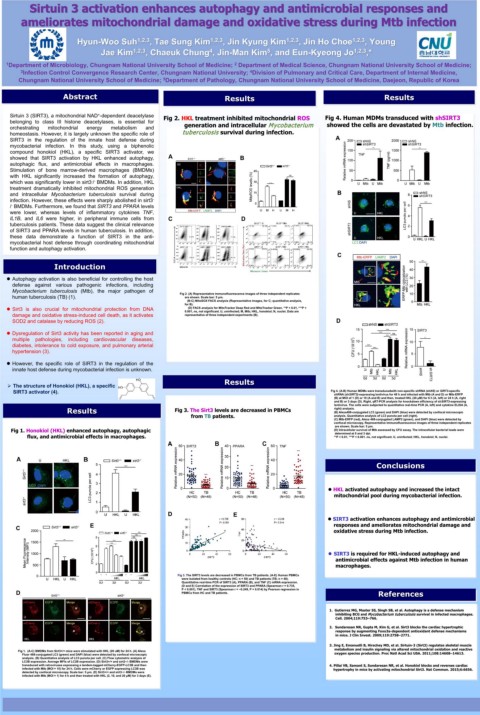
SIRT3 activator (4). Fig 4. (A-B) Human MDMs were transducedwith non-specific shRNA (shNS) or SIRT3-specific
shRNA (shSIRT3)-expressing lentivirus for 48 h and infected with Mtb (A and D) or Mtb-ERFP
(B) at MOI of 1 (D) or 10 (A and B) and then, treated HKL (20 μM) for 6 h (A, left) or 24 h (A, right
and B) or 3 days (D). Right, qRT-PCR analysis for knockdown efficiency of shSIRT3-expressing
lentivirus. The cells were subjected to quantitative real-time PCR (A, left) and cytokine ELISA (A,
Results Fig 3. The Sirt3 levels are decreased in PBMCs (B) Alexa488-conjugated LC3 (green) and DAPI (blue) were detected by confocal microscopic
right) analysis.
from TB patients. analysis. Quantitative analysis of LC3 puncta per cell (right).
(C) Mtb-ERFP (red), Alexa 488-conjugated LAMP2 (green), and DAPI (blue) were detected by
confocal microscopy. Representative immunofluorescence images of three independent replicates
are shown. Scale bar: 5 μm.
Fig 1. Honokiol (HKL) enhanced autophagy, autophagic (D) Intracellular survival of Mtb assessed by CFU assay. The intracellular bacterial loads were
determined at 0 and 3 dpi.
flux, and antimicrobial effects in macrophages. **P < 0.01, ***P < 0.001. ns, not significant. U, uninfected; HKL, honokiol; N, nuclei.
Conclusions
HKL activated autophagy and increased the intact
mitochondrial pool during mycobacterial infection.
D E
SIRT3 activation enhances autophagy and antimicrobial
C E responses and ameliorates mitochondrial damage and
oxidative stress during Mtb infection.
SIRT3 is required for HKL-induced autophagy and
antimicrobial effects against Mtb infection in human
macrophages.
Fig 3. The SIRT3 levels are decreased in PBMCs from TB patients. (A-E) Human PBMCs
were isolated from healthy controls (HC; n = 50) and TB patients (TB; n = 48).
Quantitative real-time PCR of SIRT3 (A), PPARA (B), and TNF (C) mRNA expression.
(D and E) Correlation of the expression of SIRT3 and PPARA (Spearman r = 0.735,
P < 0.001), TNF and SIRT3 (Spearman r = −0.249, P = 0.014) by Pearson regression in
D PBMCs from HC and TB patients. References
1. Gutierrez MG, Master SS, Singh SB, et al. Autophagy is a defense mechanism
inhibiting BCG and Mycobacterium tuberculosis survival in infected macrophages.
Cell. 2004;119:753–766.
2. Sundaresan NR, Gupta M, Kim G, et al. Sirt3 blocks the cardiac hypertrophic
response by augmenting Foxo3a-dependent antioxidant defense mechanisms
in mice. J Clin Invest. 2009;119:2758–2771.
3. Jing E, Emanuelli B, Hirschey MD, et al. Sirtuin-3 (Sirt3) regulates skeletal muscle
metabolism and insulin signaling via altered mitochondrial oxidation and reactive
Fig 1. (A-C) BMDMs from Sirt3+/+ mice were stimulated with HKL (20 uM) for 24 h. (A) Alexa
Fluor 488-conjugated LC3 (green) and DAPI (blue) were detected by confocal microscopic oxygen species production. Proc Natl Acad Sci USA. 2011;108:14608–14613.
analysis. (B) Quantitative analysis of LC3 puncta per cell. (C) Flow cytometric analysis of
LC3B expression. Average MFIs of LC3B expression. (D) Sirt3+/+ and sirt3−/- BMDMs were
transduced with retroviruses expressing a tandem-tagged mCherry-EGFP-LC3B and then 4. Pillai VB, Samant S, Sundaresan NR, et al. Honokiol blocks and reverses cardiac
infected with Mtb (MOI = 10) for 24 h. Cells were mCherry or EGFP expressing LC3B was hypertrophy in mice by activating mitochondrial Sirt3. Nat Commun. 2015;6:6656.
detected by confocal microscopy. Scale bar: 5 μm. (E) Sirt3+/+ and sirt3−/- BMDMs were
infected with Mtb (MOI = 1) for 4 h and then treated with HKL (2, 10, and 20 μM) for 3 days (E).