
Page 25 - M. Immunology
P. 25
Role of mitochondria in Mycobacterium tuberculosis infected macrophages
Junghwan Lee , Ji-Ae Choi , Soo-Na Cho , Sang-Hun Son , Doan Tam Nguyen , Seong-Ahn Lee 1,2 and Chang-Hwa Song 1,2*
1,2
1,2
1,2
1,2
1,2
2
1
Department of Medical Science , and Department of Microbiology , College of Medicine, Chungnam National University, 266 Munhwa-ro,
Jung-gu, Daejeon 35015, South Korea
BACKGROUND AIM
Many intracellular bacteria use host organelles such as the mitochondria, Golgi, Recent studies have shown that bacterial infection modulates mitochondrial
and endoplasmic reticulum (ER). Mitochondria are highly dynamic organelles that dynamics in various ways to interfere with apoptosis. Previously, we reported
build the large interconnected intracellular networks responsible for cellular that endoplasmic reticulum (ER) stress-mediated apoptosis is important
metabolism, signaling, and death. Mitochondrial dynamics are controlled by their defense mechanisms to anti-mycobacteria. However, relationship between
fusion and fission. Mitochondrial fusion involves the outer mitochondrial membrane ER and mitochondria has not been fully understood in Mtb-infected
GTPases mitofusin 1/2 (MFN1/2), and inner membrane GTPase optic atrophy 1 macrophages. Therefore, we investigated whether Mtb-induced ER stress
(OPA1). Mitochondrial fission requires mitochondrial fission 1 protein (FIS1) and causes mitochondrial network fragmentation in murine macrophages.
GTPase dynamin-related protein 1 (DRP1), but the molecular details of fusion and
fission are unknown. Mitochondrial fission seems to be an early stage of apoptosis.
Additionally, mitochondrial dynamics function as signals to induce the innate
immune response during viral infection. However, the role of mitochondrial METHODS
dynamics during infection is unclear. In mammalian cells, the E3 ubiquitin ligase
Parkin regulates mitochondrial dynamics by promoting proteasome-dependent Bone marrow-derived macrophages (BMDMs) were isolated and
degradation of MFN1/2. Parkin is involved in the innate immune response to differentiated by growth for 4 days in 25 ng/ml macrophage colony-
Mycobacterium tuberculosis (Mtb) and Mycobacterium leprae infection by stimulating factor containing DMEM supplemented with 10 % FBS. The
promoting ubiquitin-mediated autophagy of mycobacteria and inhibiting degree of mitochondrial fragmentation was acquired using a confocal laser-
mycobacterial replication in macrophages. ER stress is implicated in the response scanning microscope. Production of ER stress molecules and mitochondrial
to Mtb and regulates apoptosis in the presence of a variety of pathogens. fusion/fission makers was analyzed by western blotting. Cell death was
However, the relationship between ER stress-mediated apoptosis and assessed using an Annexin V/PI staining kit.
mitochondrial dysfunction is unclear.
RESULTS
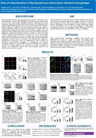
Figure. 1. Mtb increases mitochondrial
dysfunction in macrophages.
Bone marrow-derived macrophages (BMDMs)
were infected with Rv or Ra (MOI = 1) for 3 h
and incubated for 24 and 48 h. Mitochondrial
membrane potential in BMDMs was assessed
by JC-1 staining. Fluorescence of JC-1 was
detected at an excitation wavelength of 488 nm
and an emission wavelength of 530 nm by (A)
confocal microscopy or (C) flow cytometry. (B)
Quantification of the red-to-green ratio in (A). (D)
Oxygen consumption rate after sequential
treatment with oligomycin, CCCP, and rotenone.
(E) ATP production in Mtb-infected BMDMs.
ATP production rate was calculated from OCR
measured in the XF24 Extracellular Flux
Analyzer. ATP production rate was calculated Figure. 2. Mtb induces mitochondrial fragmentation in macrophages.
from OCR measured in the XF24 Extracellular (A) BMDMs were infected with RFP-labeled Rv or Ra (red), incubated for 48 h, stained with Tom20 to detect
Flux Analyzer. Data are means ± SD of three mitochondria (green) and DAPI to visualize nuclei (blue), and visualized using confocal microscopy. (B)
independent experiments. *P < 0.05, **P < 0.01. Quantification of the mitochondrial morphology (aspect ratio; left, and circularity; right) in (A). (C and D)
Cells were harvested and subjected to western blotting for mitochondrial fusion/fission factors. (E) BMDMs
were infected with Rv or Ra (MOI=1 to 5) for 3 h, and then incubated 48 h.
Figure. 5. Mtb-induced Parkin regulates
MFN2 production via the ubiquitin-
proteasome system.
(A) BMDMs were infected with Rv or Ra for
3 h. The cell lysates were subjected to
western blotting for ubiquitin. (B) BMDMs in
the presence or absence of ER stress
inhibitors or (C and D) MG132 for 1 h,
before infection with Rv or Ra. At 48 h after
Mtb infection, western blotting for ubiquitin,
Parkin, and MFN2 was performed. β-actin
was used as the loading control.
Figure. 6. MFN2 regulates intracellular
survival of Mtb.
BMDMs were transfected with siRNA. (A)
Apoptosis and (B and C) intracellular
survival of siRNA-treated BMDMs infected
with Rv or Ra for 24 and 48 h. (D) BMDMs
were transfected with pcDNA3.1-MFN2,
infected with Mtb (MOI = 1) for 24 and 48
h, and apoptosis was analyzed by flow
cytometry. (E and F) MFN2-
Figure. 3. Mtb infection decreases MFN2 overexpressing cells were infected with Rv
production via Parkin induction. Figure. 4. Mtb-induced ER stress modulates or Ra (MOI = 1) for 24 and 48 h, and
(A and B) Raw264.7 cells or BMDMs were infected MFN2 production in macrophages. intracellular survival was assayed by
with Rv or Ra (MOI = 1) for 3 h. After washing, the (A and B) Raw264.7 cells and BMDMs were enumerating CFU. Intracellular survival of
cells were incubated for 24 and 48 h. MFN2 and infected with Mtb at a MOI of 1 for 3 h. BMDMs Mtb was expressed as the change (n-fold)
Parkin were detected by western blotting. (C) were pretreated with (C) tunicamycin (Tm, 500 in the bacterial number at a given time
Representative confocal images of Mtb-infected ng/mL), (D) 4-phenylbutyric acid (4-PBA, 10 mM), point relative to the initial number of
cells. DNA was stained with DAPI (blue) and MFN2 or (E) ER stress inhibitors (10 µM GSK2606414, invasive bacteria. Data are means ± SD of
(green). BMDMs were transfected with siNegative 5 µM IREstatin, 500 µM AEBSF) for 1 h and three independent experiments. *P < 0.05,
(200 nM) or siParkin (200 nM), and infected with infected with Mtb for 3 h. At 48 h after Mtb **P < 0.01.
Rv or Ra (MOI = 1; 48 h). In siRNA-transfected infection, western blotting for Parkin, MFN2,
BMDMs, we measured the (D and E) expression of CHOP, and β-actin was performed.
MFN2 and Parkin using western blotting.
CONCLUSION REFERENCES ACKNOWLEDGEMENTS
The work was supported by the research fund of Chungnam National
Taken together, the reduced level of MFN2 ■ Sci Signal 2009, 2, ra47, doi:10.1126/scisignal.2000287. University and the Brain Korea 21 PLUS Project for Medical Science,
Chungnam National University.
production is important for anti-mycobacterial ■ EMBO Rep 2010, 11, 133-138, doi:10.1038/embor.2009.258.
function of macrophages. Mtb-induced ER stress ■ PNAS, 108, 3612-3617, doi:10.1073/pnas.1100126108.
is associated with disruption of mitochondrial Contact information
structural dynamics in macrophages. ■ Nature 2013, 501, 512-516, doi:10.1038/nature12566. E-mail: asrai1509@gmail.com