
Page 21 - M. Immunology
P. 21
Exclusive traits of neutrophils in NLRP3 inflammasome signaling
contribute to prolonged inflammation
Seunghwan Son, Do-Wan Shim, Inhwa Hwang, Je-Wook Yu
Department of Microbiology, BK 21 PLUS project for Medical Science, Yonsei University College of Medicine, Seoul, Korea
ABSTRACT A B
Deregulated inflammasome signaling contributes to diverse chronic inflammatory diseases,
but the specific cell type responsible for sustained inflammasome response remains elusive.
Here we present evidences that neutrophils can facilitate sustained inflammasome-
response and IL-1β secretion. We showed neutrophils resist to undergo cell death,
including pyroptosis upon inflammasome activation, unlike macrophages. Consistently,
intraperitoneal LPS administration caused a marked NLRP3-dependent pyroptosis of
macrophages, but much less of neutrophils in the peritoneal lavage of mice. Furthermore,
NLRP3 inflammasome activated neutrophils preserved their functional integrity,
encompassing phagocytosis and degranulation. Intriguingly, pretreatment with danger
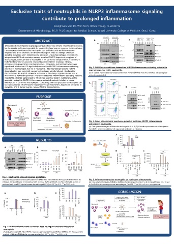
signals-rich medium or ATP significantly desensitized NLRP3 inflammasome-activating Fig. 3. DAMP-rich conditions desensitize NLRP3 inflammasome activating potential in
potential of macrophages, but not of neutrophils. This macrophage-specific NLRP3
desensitization was potentially caused by the danger signals-induced mitochondrial macrophages, but not in neutrophils.
depolarization. Neutrophils showed a resistance to this danger signals-induced loss of (A, B) Cultural supernatants and soluble lysates from BMNs or BMDMs were immunoblotted with appropriate
mitochondrial membrane potential. With these sustained inflammasome-activating capacity, antibodies as indicated.
neutrophils exhibited a prolonged IL-1β-oriented secretion pattern. Additionally, unlike
apoptotic neutrophils, NLRP3 inflammasome activated neutrophils failed to induce
efferocytosis in peritoneal macrophages. Collectively, our data propose that neutrophils can A
facilitate sustained inflammasome response through mitochondria-dependent resistance to
pyroptosis and to danger signals-induced NLRP3 desensitization. B C
PURPOSE
Background
Fig. 4. Intact mitochondrial membrane potential facilitates NLRP3 inflammasome
activation in neutrophils.
(A) Flow cytometric analysis of BMNs after staining with JC-1. (B, C) Cultural supernatants and soluble lysates
from BMNs were immunoblotted with appropriate antibodies as indicated.
A
RESULTS
A B C
B C
C D
Fig. 1. Neutrophils showed impaired pyroptosis.
(A) Cultural supernatants and soluble lysates from BMNs were immunoblotted with appropriate antibodies as Fig. 5. Inflammasome-active neutrophils do not induce efferocytosis.
indicated. (B) Quantification of extracellular LDH of mouse BMNs or BMDMs. (C) Flow cytometric analysis of (A) Flow cytometric analysis of BMNs or BMDMs after staining with annexin V. (B, C) Quantification of IL-10 and
peritoneal cells from LPS injected mouse after staining with PI (*p<0.05, **p< 0.01, ***p<0.001, n=3). IL-6 mRNA level in mouse peritoneal macrophages. (*p<0.05, ***p<0.001, n=3, n.s., not significant).
CONCLUSION
A B C
Fig. 2. NLRP3 inflammasome activation does not impair functional integrity of
neutrophils.
(A, B) Quantification of IL-1β or MMP9 in culture supernatants of mouse BMNs or BMDMs. (C) Flow cytometric
analysis of BMNs or BMDMs after zymosan uptake (*p<0.05, **p< 0.01, ***p<0.001, n=3).