
Page 19 - M. Immunology
P. 19
Microphthalmia-associated transcription factor (MITF) regulates function of
myeloid-derived suppressor cells in tumor microenvironment
1
Haesun Park , Aram Lee , Jihyun Lim , and Jong-Seok Lim 1,2
1
1
2
1 Department of Biological Science, Research Institute of Women’s Health and Cellular Heterogeneity Research Center, Sookmyung Women’s
University, Seoul 04310, Republic of Korea
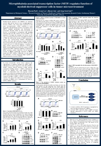
Abstract Figure 2. MITF expression increases in BM-MDSCs in tumor Figure 4. Increase of MITF expression induces the functional
microenvironment. activity of BM-MDSCs.
Myeloid cells were cultured in RPMI 1640 medium containing 10 The myeloid cells from the femurs of Balb/c were cultured with RPMI
Myeloid-derived suppressor cells (MDSCs) are immature ng/ml GM-CSF in the absence or presence of TCCM. On day 5, cells 1640 containing 10 ng/ml GM-CSF. The indicated concentration of
IBMX was added to cells. After 5 days, the cells were harvested for
myeloid cells and are known to have immunosuppressive were harvested for analysis. The IL-10, iNOS and MITF mRNA levels analysis. The cells were stained with anti-CD11b and anti-Gr-1 specific
functions. MDSCs, one of the major components of the in BM-MDSCs were measured by quantitative real-time PCR (A-C). antibodies for being analyzed by flow cytometry (A). The protein levels
The MITF, ARG1, p-STAT3 and STAT3 protein levels in BM-
tumor microenvironment (TME), contribute to tumor MDSCs were confirmed by western blot analysis (D). **p<0.01, in BM-MDSCs were confirmed by western blot analysis (B, E, F). The
progression. Microphthalmia-associated transcription factor ***p<0.001. IL-10 and iNOS mRNA levels in BM-MDSCs were measured by
quantitative real-time PCR (C, D). *p<0.05
(MITF) modulates proliferation and development of
melanocytes. Recently, MITF has been evaluated for its
function in development of non-pigment cells including
osteoclasts and mast cells. However, the role of MITF in the
regulation of immune cells remains to be elusive. In this
study, we investigated the functional role of MITF in MDSCs
in TME. We observed the increase of MITF expression in
murine bone marrow-derived MDSCs (BM-MDSCs)
cultured with tumor cell conditioned medium (TCCM). It
was accompanied by the up-regulation of MDSC activation
markers, such as IL-10, iNOS and arginase 1. In addition, an
MITF inhibitor suppressed the expression of MITF and
MDSC activation markers. In contrast, the increase of MITF
expression by IBMX induced up-regulation of expression of
MDSC activation markers. Especially, HIF-1α regulating
function of MDSCs significantly decreased in MDSCs
treated with MITF inhibitor. Collectively, our data suggest
that modulation of MITF expression might regulate the
immune suppressive function of MDSCs.
Introduction
In tumor environment, various factors interrupt the
differentiation of immature myeloid cells (IMCs). IMCs Figure 5. MITF upregulates the activation markers of BM-MDSCs
differentiate into immune suppressive cells, such as MDSCs. via HIF-1α
MDSCs suppress immune responses through multiple Bone marrow cells were obtained from the femurs of Balb/c mice. After
mechanism including arginase 1 (ARG1), interleukin-10 red blood cells were removed, myeloid cells were cultured in RPMI
(IL-10) and inducible nitric oxide synthase (iNOS). 1640 supplemented with 10% heat-inactivated FBS and 10 ng/ml GM-
MITF is a transcription factor that regulates melanin CSF, and treated with TCCM, ML-329, IBMX or 2-ME2. After 3 days,
fresh medium with recombinant GM-CSF was added. The cells were
synthesis and melanocyte development. Besides melanocyte, harvested on day 5 for analysis. The protein levels were confirmed by
MITF controls differentiation of osteoclasts and mast cells. western blot (A, B, E, F). HIF1A mRNA levels were measured by
However, the effect of MITF in MDSCs has been poorly quantitative real-time PCR (C, D). *p<0.05, ***p<0.001.
explored. The aims of this study are to investigate MITF
expression in MDSCs and to demonstrate the effect of Conclusion
MITF on MDSC activation.
Figure 3. The expressions of MDSC activation marker are
Results decreased by MITF inhibitor.
In bone marrow cells from the Balb/c mice femurs, red blood cells
were removed, and then lymphocytes were depleted using two
antibodies. The remaining myeloid cells were cultured RPMI 1640
medium containing 10 ng/ml GM-CSF, 30% TCCM or ML-329. After
5 days, cells were harvested for analysis. To confirm the population of
BM-MDSCs, cells were stained with specific antibodies against CD11b
and Gr-1. The stained cells were measured by flow cytometry (A). The
mRNA expression levels of harvested BM-MDSCs were measured by
quantitative RT-PCR for mRNA level (B, E, F), and western blot for
protein expression levels (C, D). *p<0.05, **p<0.01, ***p<0.001.
1. In tumor conditions, MITF expression is upregulated by
tumor-secreted various factors in MDSCs.
2. Increased MITF expression induces MDSC activation
Figure 1. Generation of BM-MDSCs in vitro.. throught upregulation of IL-10, iNOS and ARG1
Bone marrow cells were obtained from the femurs of Balb/c mice. After expression..
red blood cells were removed, lymphocytes were depleted using two
monoclonal antibodies. Next, myeloid cells were cultured in RPMI
1640 supplemented with 10% heat-inactivated FBS and 10 ng/ml GM- References
CSF in the absence or presence of TCCM. The cells were collected on
day 5 for analysis. The samples were analyzed by flow cytometry. 1. Busca, R., et al., Hypoxia-Inducible Factor 1{Alpha} is a
New Target of Microphthalmia-Associated Transcription
Factor (MITF) in Melanoma Cells. The Journal of Cell
Biology, 2005, 170(1): 49-59.
2. Corzo, C. A., et al., HIF-1alpha Regulates Function and
Differentiation of Myeloid-Derived Suppressor Cells in the
Tumor Microenvironment. The Journal of Experimental
Medicine, 2010, 207(11): 2439-2453.
3. Gabrilovich, D. I., and S. Nagaraj. Myeloid-Derived
Suppressor Cells as Regulators of the Immune System.
Nature Reviews Immunology, 2009, 9(3): 162-174.
4. Kitamura, Y., E. Morii, T. Jippo, and A. Ito. Effect of
MITF on Mast Cell Differentiation. Molecular
Immunology, 2002, 38(16-18): 1173-1176.
5. Nam, S., et al., Interferon Regulatory Factor 4 (IRF4)
Controls Myeloid-Derived Suppressor Cell (MDSC)
Differentiation and Function. Journal of Leukocyte
Biology, 2016, 100(6): 1273-1284.