
Page 11 - M. Immunology
P. 11
Positive Effect of Retnla Deficiency Against Lipopolysaccharide-Induced Septic Shock
1
Seo-Yeon An , Kyuseong Park , Jae-Hoon Choi , Mi-Ran Lee 3*
2
2
1 Department of Medical Science, Chungnam National University, Daejeon, Korea
2 Department of Life Science, College of Natural Sciences, Research Institute for Natural Sciences, Hanyang University, Seoul, Korea
3 Department of Biomedical Laboratory Science, Jungwon University, Chungbuk, Korea
BACKGROUND AIM
Sepsis is a life-threatening condition caused by a dysregulated host inflammatory response to various infections, In this study, we investigated the effects of Retnla in endotoxemia-
including bacterial, fungal, viral, and protozoan infection, which can lead to tissue damage, organ failure, and death. induced septic responses. To explore the effects of Retnla on
Systemic inflammatory response syndrome is the hallmark sign of sepsis and mediated by an initial intense pro- inflammation in vivo, we employed a lipopolysaccharide (LPS)-induced
inflammatory responses or cytokine storm. Inducible nitric oxide synthase is a key inflammatory mediator of sepsis and its murine sepsis model and injected LPS (10 mg/kg; Escherichia coli
product nitric oxides contribute to sepsis-mediated organ dysfunction. O111:B4) intraperitoneally into mice in the presence or absence of
Resistin-like molecule alpha (Retnla) is a cysteine-rich secreted protein and has been found in bronchoalveolar lavage Retnla. Retnla deficiency reverse the reduced cell viability of
fluid of ovalbumin-induced asthmatic mice. It plays a role as a regulator of T helper 2-driven inflammation and regulates RAW264.7 and suppressed the expression levels of IL-1b and iNOS in
the pathogenesis of inflammatory diseases such as parasitic diseases and asthma. However, the role of Retnla in LPS-activated RAW264.7 cells.
inflammatory responses against bacterial infection is unknown.
METHODS
Generation of sepsis model mice: LPS (10 mg/kg) was intraperitoneally injected to both wild-type and Retnla-deficient mice. The mice were sacrificed on 0, 6, 12, 24 h after LPS injection.
Western blot : Blood samples were collected by retro-orbital sinus puncture and were immediately centrifuged at 1,500 g for 20 min to separate serum. For Retnla detection, the serum was filtered through a 100,000-Dalton cut-off Amicon filter device
(Millipore Corporation), and 40 ml was analysed by 4–20% SDS–PAGE.
RNA isolation and quantitative real-time PCR : Total RNA from Raw264.7 cells was extracted using the Trizol reagent (5 PRIME). Then, 1.5 mg RNA was quantified spectrophotometrically and reverse transcribed into complementary DNA, using the
RevertTra Ace qPCR RT master mix (TOYOBO) according to the manufacturer’s instructions. qRT–PCR was performed with a two-step using an StepOnePlus Real-Time PCR System (Applied Biosystems) and SYBR Green
PCR master mix (Bioline). The following primers were used: iNOS forward, 5’- CCC TTC AAT GGT TGG TAC ATG G-3’ and reverse, 5’-ACA TTG ATC TCC GTG ACA GCC-3’; IL-1β forward, 5’-GGA GAA CCA AGC AAC GAC AAA ATA -3’ and
reverse, 5’- TGG GGA ACT CTG CAG ACT CAA AC -3’.
MTS assay for cell viability: RAW264.7 cells were seeded in a 96-well plate at a concentration of 5× 10 4 cells/mL, and were treated with LPS (1 μg/mL) for 24 h. Cell viability was determined in the cell culture supernatants using the CellTiter 96 AQueous
One Solution Cell Proliferation Assay (MTS) and measured at 530nm by a spectrophotometer.
NO determination: The concentration of NO in mice serum and cell culture supernatant was determined as nitrite by the Griess reagent. Blood samples were collected for 6 and 20h after LPS injection for the determination of NO. RAW264.7 cells were
seeded in a 96-well plate at a concentration of 4× 10 4 cells/mL, and were treated with LPS (1 μg/mL) for 24 h.
Survival rate : The survival rate of mice was recorded every 6 h for 96 h, and Kaplan-Meier survival curves were generated using the GraphPad Prism 6 software.
Histologic examination: Kidney was fixed in a 10% neutral buffered formalin and processed for hematoxylin & eosin (H&E) stain.
RESULTS
A A A
B B B
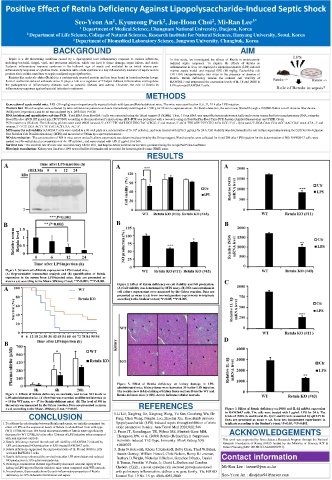
Figure 1. Serum level of Retnla expression in LPS-treated mice.
(A) Representative immunoblot analysis and (B) quantification of Retnla
expression in the serum from LPS-injected mice. Data are presented as
mean ± s.d. according to the Mann–Whitney U-test; **P<0.005; ***P<0.001. Figure 2. Effect of Retnla deficiency on cell viability and NO production. C
A (A) Cell viability was determined by MTS assay. (B) NO concentrations in
cell culture supernatant were measured by the Griess reaction. Data are
presented as mean ± s.d. from two independent experiments in triplicate
according to the Student’s t-test; *P<0.05; **P<0.005.
D
B
Figure 5. Effect of Retnla deficiency on kidney damage in LPS-
administrated mice. Kidney tissue were harvested 20 h after LPS injection.
The results show H&E-staining of kidney tissue sections from the WT and
Figure 4. Effects of Retnla deficiency on mortality and serum NO levels in Retnla-deficient mice (× 100). Arrow indicates tubular necrosis.
LPS-administrated mice. (A) Survival was recorded at different intervals (n
= 19 for WT mice, n = 17 for Retnla-deficient mice) (B) The level of NO in
the serum was measured by the Griess reaction. Data are presented as mean REFERENCES
± s.d. according to the Mann–Whitney U-test; *P<0.05. Figure 3. Effect of Retnla deficiency on iNOS and IL-1β mRNA expression
in RAW264.7 cells. The cells were treated with 1 μg/mL LPS for 24 h. The
CONCLUSION 1.Li Li1, Xingfeng He, Xingtong Wang, Yu Sun, Guosheng Wu, He levels of iNOS (A and B) and IL-1β (C and D) were measured by qRT-PCR.
Fang, Chen Wang, Pengfei Luo, Zhaofan Xia. Ruxolitinib protects
Data are presented as mean ± s.d. from two independent experiments in
1.To address the relationship between Retnla and sepsis, we initially examined the lipopolysaccharide (LPS)-induced sepsis through inhibition of nitric triplicate according to the Student’s t-test; *P<0.05; **P<0.005.
effect of LPS on the expression levels of Retnla in whole blood from wild-type oxide production in mice. Ann Transl Med 2020;8(8):546
(WT) C57BL/6J mice. We found that serum levels of Retnla were significantly 2. Pesce JT, Ramalingam TR, Wilson MS, Mentink-Kane MM, ACKNOWLEDGEMENTS
decreased in WT C57BL/6J mice after 12 times of LPS injection when compared Thompson RW, et al. (2009) Retnla (Relma/Fizz1) Suppresses
with non-injected controls. This work was supported by Basic Science Research Program through the National
2. Retnla deficiency reversed the reduced cell viability of RAW264.7 induced by Helminth-Induced Th2-Type. Immunity. PLoS Pathog 5(4): Research Foundation of Korea (NRF) funded by the Ministry of Science, ICT &
LPS and decreased NO production in LPS-treated RAW264.7 cells. e1000393 Future Planning (NRF-2015R1C1A2A01053571).
3. Retnla deficiency suppressed the expression levels of IL-1b and iNOS in LPS- 3. Ilona N.Holcomb, Rhona C.Kabakoff, Betty Chan, Thad W.Baker,
activated RAW264.7 cells. Austin Gurney, William Henzel, Chris Nelson, Henry B.Lowman,
4. Retnla deficiency enhanced the survival rate after LPS stimulation and reduced Barbara D.Wright, Nicholas J.Skelton, Gretchen Dfrantz, Daniel Contact information
NO production in LPS-administrated mice.
5. Histologic examination showed significantly decreased necrotic injury of the B.Tumas, Franklin V.Peale, Jr, David L.Shelton and Caroline
kidney in LPS-injected Retnla-deficient mice when compared with WT controls. Chebert. FIZZ1, a novel cysteine-rich secreted protein associated Mi-Ran Lee : leemr@jwu.ac.kr
6. In conclusion, these results show the anti-inflammatory properties of Retnla with pulmonary inflammation, defines a ne gene family. The EMBO
deficiency on LPS-induced inflammation and sepsis. Journal Vol. 19 No. 15 pp. 4046-4055,2000 Seo-Yeon An : dkstjdus94@naver.com