
Page 1 - M. Immunology
P. 1
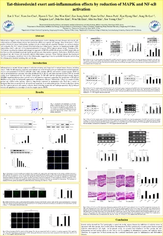
Tat-thioredoxin1 exert anti-inflammation effects by reduction of MAPK and NF-κB
                                                 activation


                         1
                                    1
             1
                                                                                                      1,3
      Eun Ji Yeo , Yeon Joo Choi , Hyeon Ji Yeo , Dae Won Kim , Eun Jeong Sohn , Hyun Ju Cha , Jinseu Park , Kyu Hyung Han , Sung Ho Lee ,
                                                                      1
                                                                               1
                                                            1
                                               2
                                                                                            1
                           Sunghou Lee , Duk-Soo Kim , Won Sik Eum , Min Jea Shin , Soo Young Choi 1,*
                                               5
                                                                     1
                                                          1
                                    4
                           1 Department of Biomedical Science and Research Institute of Bioscience and Biotechnology, Hallym University, Chuncheon 24252, Korea.
                   2 Department of Biochemistry and Molecular Biology, Research Institute of Oral Sciences, College of Dentistry, Gangneung-Wonju National University, Gangneung 25457, Korea.
                                           3 Genesen Inc., Teheran-ro, Gangnam-gu, Seoul 06181, Korea.
               4 Department of Green Chemical Engineering, Sangmyung University, Cheonan 31066, Korea.  5 Department of Anatomy, College of Medicine, Soonchunhyang University, Cheonan-Si 31538, Korea.
                        Abstract
                                                          A                                     C
  Inflammation triggers many interconnected pathophysiological events including neuronal diseases and several anti-  B
  inflammatory and anti-oxidant proteins have protective effects on neuronal cell damage. Since thioredoxin 1 (Trx1)
  protein is known to reduce cell death by regulation of anti-oxidant and anti-apoptotic functions, we explored whether
  cell permeable Tat-Trx1 protein showed beneficial influences inflammatory response in lipopolysaccharide (LPS)-
  induced Raw 264.7 cells and 12-O-tetradecanoylphorbol-13-acetate (TPA)-induced animal model. Transduced Tat-
  Trx1 protein significantly reduced cell damages by regulation of MAPK and NF-κB activation in LPS-treated cells and
  this fusion protein modulated apoptosis signaling pathway and inflammatory responses. Furthermore, Tat-Trx1 protein
  transduced into skin tissues and markedly reduced inflammatory responses. These findings indicate that transduced
  Tat-Trx1 protein paly a beneficial roles in the inflammation, suggesting it may be a potential therapeutic protein drug
  for inflammatory disorders including skin and dry eye.
                                                        Fig. 5. Effect of Tat-Trx1 protein against LPS-induced NF-κB, MAPK and Akt expression in Raw264.7 cells. One-hour pretreatment of Raw264.7 cells with Tat-
                                                        Trx1 protein (1 μM) or Trx1 protein was followed by treatment with LPS (1 μg/ml) for 120 min (p65), 30 min (MAPK) and 60min (Akt) respectively. The
                       Introduction                     expression levels of p65 (A), p38, JNK, ERK (B) and Akt (C) were determined by Western blot analysis and the band intensity was measured by densitometer. * P <
                                                        0.05 compared with LPS-treated cells.
  Inflammation is a natural defense response to infection or injury, and it may lead to various human diseases, including
  cancer. Under the inflammatory responses, macrophages are activated and secrete pro-inflammatory mediator proteins,
  such as cyclooxygenase-2 (COX-2), inducible nitric oxide synthase (iNOS), and reactive oxygen species (ROS) as
  well as pro-inflammatory cytokines, including interleukin (IL)-6, IL-1β, and tumor necrosis factor-α (TNF-α). Several  A  B
  studies have demonstrated that the nuclear factor-kappa B (NF-κB) and the mitogen-activated protein kinases
  (MAPKs) signaling pathways play a pivotal role in inflammatory responses, suggesting that modulation of NF-κB and
  MAPKs is a key point for therapeutic approaches to inflammatory diseases. Thioredoxin 1 (Trx1) is a small (12 kDa)
  protein and cellular redox enzyme, which is ubiquitously expressed in mammalian cells. Trx1 has a variety of
  biological functions in regulation of cell growth and apoptosis as an antioxidant protein. Several studies have shown
  that Trx1 protein protects cells against oxidative stress including superoxide and hydrogen peroxide (H 2 O 2 )-induced
  toxicity and apoptosis as a scavenger of reactive oxygen species (ROS).
                                                        Fig. 6. Effect of Tat-Trx1 protein against LPS-induced Bcl-2, Bax and cleaved caspase-3 expression in Raw264.7 cells. One-hour pretreatment of Raw264.7 cells
                                                        with Tat-Trx1 protein (1 μM) or Trx1 protein was followed by treatment with LPS (1 μg/ml) for 120 min (Bcl-2, Bax), and 180 min (cleaved caspase-3)
                         Results                        respectively. The expression levels of Bcl-2, Bax (A) and cleaved caspase-3 (B) were determined by Western blot analysis and the band intensity was measured by
                                                        densitometer. * P < 0.05 compared with LPS-treated cells.
                    B
                                                       A                         B
     A                            Fig. 1. Construction and purification of Tat-Trx1
                                  protein. Constructed map of Tat-Trx1 based on the
                                  pET15-b vector and diagrams of the expressed Tat-
                                  Trx1 proteins (A). Purified recombinant Tat-Trx1 and
                                  Trx1 proteins were identified by 15% SDS-PAGE and
                                  detected by Western blot analysis using an anti-
                                  histidine antibody (B).
    A                        B                          Fig. 7. Effect of Tat-Trx1 protein against LPS-induced inflammatory responses in Raw264.7 cells. One-hour pretreatment of Raw264.7 cells with Tat-Trx1 protein
                                                        (1 μM) or Trx1 protein was followed by treatment with LPS (1 μg/ml). The expression levels of iNOS and COX-2 (A) protein were analyzed by Western blotting.
                                                        Total RNA was extracted from the cells/ We analyzed cytokines (IL-1β, IL-6 and TNF-α) and GAPDH mRNA by RT-PCR using specific indicated primers (B).
                                                        The band intensity was measured by densitometer. * P < 0.05 compared with LPS-treated cells.
                                                               A
    C                        D
                                                          B
   Fig. 2. Transduction of Tat-Trx1 proteins into Raw264.7 cells. Raw264.7 cell culture media were treated with Tat-Trx1 protein at different doses (0.1-1
   μM) or with the Trx1 protein for 1 h (A). The cell culture media were treated with Tat-Trx1 protein (1 μM) or Trx1 protein for different time periods (15-60
   min) (B). Then, transduction of Tat-Trx1 protein was measured by Western blotting and the intensity of the bands was measured by a densitometer.
   Intracellular stability of transduced Tat-Trx1 protein. Raw264.7 cell culture media were incubated for 12 h after transduction of Tat-Trx1 protein for 1 h (C).
   Transduction of Tat-Trx1 protein was measured by Western blotting and the intensity of the bands was measured by a densitometer. The localization of
   transduced Tat-Trx1 protein was examined by confocal fluorescence microscopy (D). Scale bar = 50 μm.


   Fig. 3. Effects of Tat-Trx1 protein on LPS-induced ROS production. Treatment with Tat-Trx1 protein (1 μM) and Trx1 protein was followed by 1 h treatment
   with LPS (1 μg/ml). Intracellular ROS levels were measured by DCF-DA staining and fluorescence intensity was measured by ELISA plate reader; scale bar
   = 50 μm. * P < 0.05 compared with LPS-treated cells.
                                                        Fig. 8. Effects of Tat-Trx1 on TPA-induced pro-inflammatory mediators (iNOS and COX-2) and cytokine (IL-1β, IL-6 and TNF-α) expression in mice ears (A).
                                                        Mice were stimulated with TPA (1 μg/ear) after which Tat-Trx1 (10 μg) was topically applied to mice ear for 3 days. After total RNA was extracted from ear
                                                        biopsies, pro-inflammatory mediator and cytokine levels were determined by RT-PCR. The band intensity was measured by densitometer. Effects of Tat-Trx1 on
                                                        TPA-induced mice ear edema. Ears of mice were treated with TPA (1 μg/ear) once a day for 3 days. Tat-Trx1 protein (10 μg) was topically applied to mice ears 1
                                                        h prior to TPA exposure over 3 days. Protective effects of Tat-Trx1 were confirmed by hematoxylin and eosin staining as well as changes in ear weight and ear
                                                        thickness in a TPA-induced mice ear edema model (B). Scale bar = 50 μm. * P < 0.05 and ** P < 0.01 compared with TPA-treated mice.
                                                                              Conclusion
                                                       Several studies showed that Trx1 protein play a central role in the various diseases by inhibition of oxidative stress. In
                                                       agreement with other groups, these results indicate that transduced Tat-Trx1 protein plays an anti-oxidant function against
                                                       oxidative stress-induced cell death.  In the present study, we revealed that transduced Tat-Trx1 protein has anti-
                                                       inflammatory and anti-oxidant effects in vitro and in vivo by regulating of inflammatory response and oxidative stress.
   Fig. 4. Effect of transduced Tat-Trx1 protein on DNA damage. The cells were treated with Tat-Trx1 (1 μM) for 1 h, and then exposed to LPS (1 μg/ml) for
   12 h. Then, DNA fragmentation was detected by TUNEL staining and fluorescence intensity levels were measured by ELISA plate reader. Scale bar = 50  Therefore, we suggest that Tat-Trx1 protein may be a potential therapeutic agent for inflammation and ROS-relative
   μm. * P < 0.05 compared with LPS-treated cells.     diseases.