
Page 9 - G. Cell differentiation. division. and death
P. 9
CHIP negatively regulates necroptosis via ubiquitylation-meidated
lysosomal degradation of RIPK1 and RIPK3.
Choong-Sil Lee¹,²,# Jinho Seo³,# Eun-Woo Lee⁴,# Jaewhan Song¹,²,*
¹Department of Biochemistry, College of Life Science and Biotechnology, Yonsei University, Seoul 120-749, Korea, ²Interdisciplinary Program of Integrated OMICS for Biomedical Science,
WCU, Yonsei University, Seoul Korea, Korea, ³Environmental Disease Research Center, Korea Research Institute of Bioscience and Biotechnology (KRIBB), Daejeon 34141, Korea,
⁴Metabolic Regulation Research Center, Korea Research Institute of Bioscience and Biotechnology (KRIBB), Daejeon 34141, Korea * Co-Coressponding Author # Co-first Author
BACKGROUND AIM
TNFα signaling is capable of inducing cell growth, survival, inflammation The mechanisms by which TNFα activation leads to either apoptosis or
and cell death. It determines the outcome depending on complex necroptosis have been analysed comprehensively in biochemical and genetic
external and internal context of the cells. Several checkpoints of TNFα contexts. However, the regulatory mechanism at the protein level of these
signaling including kinases, E3 ligases, deubiquitylases and others have factors is yet to be uncovered. Here we report that CHIP (STUB1) regulates
RIPK1 and RIPK3 protein stability by lysosomal degradation via its E3 ligase
been reported to regulate cell fate. activity.
METHODS
Mice : Chip -/- RIPK3 -/- DKO mice were generated from Chip -/- (from RIKEN) and Ripk3 -/- (from Genentech) mice.
Histology : 4-week-old mice were dissected to analyze thymus and small intestine. The tissues were fiexed in 4 % neutral buffered paraformaldehyde, embedded in paraffin and sliced into 3μm sections. H&E staining and TUNEL assay
were performed using standard procedure from producers.
Cell culture : MEF, 293FT, H1299 and HT-29 cell lines were cultured in DMEM or RPMI supplemented with 10% FBS and 1 % penicillin/streptomycin.
Cell treatment : To induce necroptotic cell death, 30 ng/ml TNFα, 2 μg/ml cycloheximide and 30μM z-VAD-fmk were treated for MEFs. 30 ng/ml TNFα, 5 μg/ml cycloheximide and 30 μM z-VAD-fmk were treated for HT-29. To inhibit
necroptosis, 30 μM necrostatin-1 was treated. 293FT cells were transfected with plasmid by PEI. MEF, HT-29 and H1299 cells were transfected with siRNA or plasmid by Lipofectamine 2000 or RNAiMAX.
Immunoprecipitation and Western-Blotting : Cells were lysed by a lysis buffer (50 mM Tris-HCl, pH=7.5, 150 mM NaCl, 1.0 % Triton X-100, 1 mM EDTA and 10 % glycerol, protease inhibitors). For immunoprecipitation, antibodies
against target proteins were incubated with cell lysates. protein-G-agarose bead were incubated to pulldown target proteins. For Western-blot, samples were blotted by Laemmli sample buffer and SDS-PAGE followed by transfer (Bio-
rad). Antibodies against target proteins were incubated with blots to detect proteins.
FACS : MEF and HT-29 cells treated by necroptotic treatment, collected and resuspended in annexin-V, or 7-AAD staining-buffer according to the manufacturer’s protocol. FACScan flow cytometer (BD biosience) were used to analyze
cell-death.
Cll viability assay : Cell loss was measured by the Cell Titer Glo Luminescent Cell viability Assay Kit (Promega) according to manufacturer’s protocol.
Real-tim PCR : Trizol (Takara Bio) was used to extract RNA from cells and cDNA was synthesized by reverse transcriptase (Takara Bio). Target mRNA levels were measured by SYBR Green PCR Kit (Qiagen).
Immunofluoroscent : Cells were incubated in 100 μM LysoTracker (Invitrogen) for 1 h for lysosome staining. Cells were fixed with a 10 % formalin solution and stained by antibodies to detect target proteins.
Ubiquitination assay : cells were lysed by 1 % SDS in PBS or 6 M guanidine buffer. Antibodies against target tag or Ni2+-NTA beads were incubated with lysate to pull down ubiquitin or target protein. The ubiquitination was analyzed
by Western-Blotting.
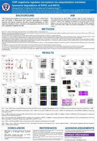
RESULTS
Figure 1 Figure 2 Figure 3
a b a b a b c d
e f g
c d
c
Figure 6
a b c
Figure 4 Figure 5
a b c d a b
d e
c d
e f g h
f g
e f
Fig 1. : Chip -/- MEFs show increased PI staining and cell loss after TNFα-induced necroptosis. Reconstitution of Chip in KO MEF rescues the sensitized cell death to the level of Chip +/+ MEF.
Fig 2. : RIPK1 and RIPK3 are increased at protein level in Chip -/- MEFs and recruited to FADD more than in Chip +/+ MEFs. The increased protein level of RIPK1 and RIPK3 is rescued by
reconsitution of Chip.
Fig 3. : CHIP overexpression decreases RIPK1 and RIPK3 level, which is dependent on E3 ligase activity of CHIP. The reduction of RIPK1 and RIPK3 by CHIP overexpression is blocked by
lysosomal inhitors E64d and pepstatin A.
Fig 4. : Overexpressed CHIP ubiquitylates RIPK1 and RIPK3. The ubiquitylation of RIPK1 and RIPK3 is increased by lysosomal inhibitors. Depletion of CHIP by siRNA decreases RIPK1 and
RIPK3 ubiquitylation. Purified CHIP directly ubiquitylates RIPK1 and RIPK3 in vitro.
Fig 5. : RIPK1 or RIPK3 and CHIP co-localize into lysosome, which is dependent on E3 ligase activity of CHIP.
Fig 6. : Ripk3 -/- Chip -/- DKO mice is rescued from Chip -/- phenotypes. The dwarfism and lethality of Chip -/- mice are restored by Ripk3 -/-. Ripk3 deletion suppresses abnormal histology of
Chip -/- thymus and small intestine.
CONCLUSION REFERENCES ACKNOWLEDGEMENTS
• CHIP (Stub1) is a negative regulator of TNF-induced Vanden Berghe et al. Regulated necrosis: the expanding network of non-apoptotic cell death This research was partially supported by the Graduate School of YONSEI
pathways. Nat. Rev. Mol. Cell Biol. 15, 135–147 (2014).
necroptosis in its E3-ligase activity dependent University Research Scholarship Grants in 2020
manner. Ahmed, S. F. et al. The chaperone-assisted E3 ligase C terminus of Hsc70-interacting protein
(CHIP) targets PTEN for proteasomal degradation. J. Biol. Chem. 287, 15996–16006 (2012).
• CHIP (Stub1) interacts with RIPK1 and RIPK3, Petrucelli, L. et al. CHIP and Hsp70 regulate tau ubiquitination, degradation and aggregation. Contact information
promoting their lysosomal degradation. Hum. Mol. Genet. 13, 703–714 (2004).
Kaiser, W. J. et al. RIP3 mediates the embryonic lethality of caspase-8-deficient mice. Nature
• Unregulated RIPK3 mediates lethality of Chip (Stub1) 471, 368–372 (2011). First author : lcs0705@yonsei.ac.kr
-/- mice. Min, J. N. et al. CHIP deficiency decreases longevity, with accelerated aging phenotypes
accompanied by altered protein quality control. Mol. Cell. Biol. 28, 4018–4025 (2008). Corresponding author : jso678@yonsei.ac.kr