
Page 7 - G. Cell differentiation. division. and death
P. 7
In vitro differentiation of Endothelial Cells from
porcine Epiblast Stem Cells
Bo-Gyeong Seo, Cheol Hwangbo , Division of Applied Life Science, College of Natural Sciences, Gyeongsang National University
†
BACKGROUND AIM
Epiblast stem cells
Pluripotent stem cells including embryonic stem cells (ESCs), epiblast stem cells (EpiSCs) and We aimed to differentiate porcine epiblast stem cells (pEpiSCs) as one of
induced iPSCs have the potential of self-renewal, and differentiate into any cell type (endoderm, pluripotent stem cells into functional vascular endothelial cells (ECs) for
mesoderm and ectoderm) in the adult body. ESCs were derived from the inner cell mass of a treatment of vascular disease. So, we have examined that differentiation
pre-implantation embryo. Since then, ESCs were able to induce successfully from primate medium induces the differentiation of pEpiSCs into vascular ECs. Also,
species. EpiSCs were derived from the late epiblast presented in a post-implantation embryo. whether the addition of VEGF to differentiation medium improves the
Vascular Endothelial Growth Factor (VEGF) efficiency of differentiation of the pEpiSCs into ECs was examined. These
VEGF in developing adult tissues is a well-established mitogen, as differentiation and survival studies have established an efficient protocol that differentiates ECs from
factors of endothelium. Additionally, VEGF has the ability to promote the proliferation of ECs. So, pEpiSCs with 2-dimensional culture.
VEGF play important roles specifically in angiogenesis and vasculogenesis process.
METHODS
Manual
Picking - Incubation at
Differentiation Medium 39℃, 5%
CO₂
‣ EBM-2
‣ EBM-2 + VEGF - Medium
‣ APEL-2 change on
Porcine Epiblast Stem Cells 0.5% gelatin coated well ‣ APEL-2 + VEGF
8 line on inactivated MEF day 2 and 5
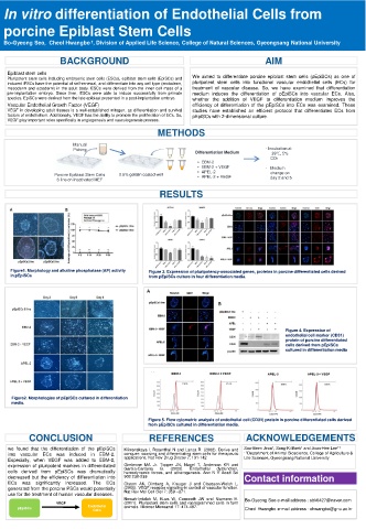
RESULTS
Figure1. Morphology and alkaline phosphatase (AP) activity Figure 3. Expression of pluripotency-associated genes, proteins in porcine differentiated cells derived
in pEpiSCs from pEpiSCs culture in four differentiation media.
Figure 4. Expression of
endothelial cell marker (CD31)
protein of porcine differentiated
cells derived from pEpiSCs
cultured in differentiation media
Figure2. Morphologies of pEpiSCs cultured in differentiation
media.
Figure 5. Flow cytometric analysis of endothelial cell (CD31) protein in porcine differentiated cells derived
from pEpiSCs cultured in differentiation media.
CONCLUSION REFERENCES ACKNOWLEDGEMENTS
we found that the differentiation of the pEpiSCs Klimanskaya I, Rosenthal N and Lanza R. (2008). Derive and Soo-Been Jeon 1 , Sang-Ki Baek 1 and Joon-Hee Lee 1†
into vascular ECs was induced in EBM-2. conquer: sourcing and differentiating stem cells for therapeutic - 1 Department of Animal Bioscience, College of Agriculture &
Especially, when VEGF was added to EBM-2, applications. Nat Rev Drug Discov 7: 131-142 Life Sciences, Gyeongsang National University
expression of pluripotent markers in differentiated Gimbrone MA Jr. Topper JN, Nagel T, Anderson KR and
Endothelial
dysfunction,
Garcia-Cardena
G.
(2000).
cells derived from pEpiSCs was dramatically hemodynamic forces, and atherogenesis. Ann N Y Acad Sci
decreased but the efficiency of differentiation into 902:230-239. Contact information
ECs was significantly increased. The ECs Olsson AK, Dimberg A, Kreuger J and Claesson-Welsh L.
generated from the porcine PSCs would potentially (2006). VEGF receptor signaling-in control of vascular function.
use for the treatment of human vascular diseases. Nat Rev Mol Cell Biol 7: 359 –371.
Nowak-Imialek M, Kues W, Carnwath JW and Niemann H. Bo-Gyeong Seo e-mail address : sbk6427@naver.com
VEGF (2011). Pluripotent stem cells and reprogrammed cells in farm
pEpiSCs Endothelial animals. Microsc Microanal 17: 474-497. Cheol Hwangbo e-mail address : chwangbo@gnu.ac.kr
Cells