
Page 5 - G. Cell differentiation. division. and death
P. 5
Diesel particulate matter 2.5 induces inflammation and mitochondrial impairment
via up-regulation of reactive oxygen species in corneal epithelial cells
4
Da Hye Kim , Hyesook Lee , Jeong-Hwan Kim , Seh-Kwang Park , Ji-Won Jeong , Mi-Young Kim , Soo-Wan Nam , Yung Hyun Choi 2,3,6 ,*
1
4
2,3
1,6
4
5
1 Department of Smart Bio-Health, Graduate School, Dong-eui University, Busan 47340,
2 Anti-Aging Research Center, Dong-eui University, Busan 47340,
3 Department of Biochemistry, Dong-eui University College of Korean Medicine, Busan 47227,
4 Research and Development Department, BGN CARE Co., Ltd., Busan 47195,
5 BGN Eye Clinic, Busan 47195,
6 Biomedical Engineering and Biotechnology Major, Division of Applied Bioengineering, College of Engineering, Dong-Eui University, Busan 47340, Republic of Korea
BACKGROUND AIM
Environmental pollution, particularly air pollution, has become a serious At present, the biggest cause and problem of air pollution is particulate matter. Particulate
issue for human health during the development of modern society. Fine PM is matter(suspended particulate matter, SPM, atmospheric aerosol particles, atmospheric
referred to as PM 2.5 (particulate matter with a diameter less than 2.5 μm), and particulate matter) is caused by various factors such as desertification, factory, exhaust
several studies have linked PM 2.5 to ocular surface diseases. Nevertheless, gas and various combustion. Particulate matter is composed of various materials such as
nitrate, ammonium, carbon compounds, sulfates, etc., which is a mixed material. We
the few studies on the biological effect and underlying mechanism of PM 2.5 on experimented with Particulate matter with diameters of less than 2.5 µm. PM 2.5 is smaller
the eye have been limited. Therefore, the aim of this study was to evaluate the than hair. PM 2.5 can already adsorb to the alveoli and reach the retina by passing through
biological effect of PM 2.5 on primary rat corneal epithelial cells (RCECs) and to the cornea.
identify the mechanism. Exposure to PM 2.5 induced cytotoxicity and DNA Rat corneal epithelial cells (RCECs) is a primary cultured cell extracted from rat. The
damage in a dose-dependent manner. The result of gene expression cornea is an organ that refracts light and helps to form an image on the retina.
microarray showed that PM 2.5 enhanced the expression of pro-inflammatory Transparency and a smooth surface are required. So the cornea is devoid of blood
mediators, such as cytokines and chemokines. We also found that up- vessels and consists of nerve cells. Recently, as air pollution caused by PM 2.5 has
regulation of the secretion of inflammatory cytokines was accompanied by increased, patients visiting conjunctivitis and dryness have been rapidly increasing. Our
activation of NF-κB and phosphorylation of p38 MAPK. It is worth noting that cornea is the most dangerous because it is the primary exposure organ to direct exposure
PM2.5 markedly increased intracellular reactive oxygen species (ROS) and to PM 2.5 . However, studies on cornea and PM 2.5 have not been actively conducted. So we
suppressed the protein expression of mitophagy regulator, such as PINK, studied how the PM 2.5 affects the cornea.
Parkin and Wee1. However, the inhibition of ROS by N-acetylcysteine METHODS
significantly suppressed the PM 2.5 -mediated cellular dysfunction including
expression and secretion of inflammatory mediators, mitochondrial Cell viability : CCK-8 assay
membrane potential loss, DNA damage and NF-κB activation. In conclusion, DNA damage : DAPI and p-γH2AX fluorescence staining
our data suggested that PM 2.5 -induced inflammation and mitochondrial
dysfunction are dependent on the ROS/NF-κB signaling pathway in RCECs. Cytokine level : mouse enzyme-linked immunosorbent assay kit
The present study provides the important evidence for understanding of Mitochondria activity: Staining with Mitotracker fluorescence
PM2.5-mediated underlying mechanism in cornea.
Keywords: Particulate matter 2.5; corneal epithelial cells; mitochondrial ROS amount : Staining with DCF-DA fluorescence
dysfunction; inflammation; reactive oxygen species Protein level : Western blotting
RESULTS
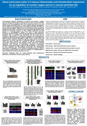
Figure 1. Effects of PM 2.5 on the cell Fig 2. Heatmap of candidate gene Figure 3. Effects of PM 2.5 on Figure 4. Effects of PM 2.5 on the NF-kB
expression using NanoString
viability in primary rat corneal nCounter® miRNA Expression Assays cytotoxicity in RCECs. induced inflammation in RCECs.
epithelial cells (RCECs). in PM 2.5 -stimulated RCECs.
Cells were treated with different concentration of PM 2.5 for Cells were treated with PM 2.5 of 100 µg/mL and 200 µg/mL for
Cells were treated with different concentration of PM 2.5
for 24 hours. (A) Cell viability was measured by CCK-8 24h. (A) PM 2.5 -induced ROS production was assessed by 24 h. (A) The production of cytokine on cell supernatant were
assay. Data are expressed as the mean ± SD (n=3). The DCFDA staining in RCEC. (B) Mitotracker staining was measured by ELIAS kits. Data are expressed as the mean ±
statistical analyses were conducted using analysis of performed to measure mitochondrial activity. (C) SD (n=3). The statistical analyses were conducted using
variance (ANOVA-Tukey’s post hoc test) between Mitophagy-related proteins were detected by Western analysis of variance (ANOVA-Tukey’s post hoc test) between
groups. *p<0.05, **p<0.01 and ***p<0.001 between blot.(D) The cells were immunofluorescence stained with groups. *p<0.05, **p<0.01 and ***p<0.001 between groups. (B)
groups. (B) The representative morphological changes anti-p-γH2AX (Ser139) antibody (red), and the nuclei were The translocation of NF-κB in the nucleus to cytosol was
of cells were taken using an inverted microscope (scale Cells were treated with 200 μg/ml of PM 2.5 for 24 h. Total counterstained with DAPI (blue). (A,B,C, Scale bar; 75 μm) immunofluorescence stained with anti-NF-κB (red) (scale bar;
bar: 75 μm). RNA was collected by collecting the cells, and 75 μm) (C) p65 detected by Western blot. (D) MAPK signaling
hybridization was performed using a reporter probe and a pathway-related protein was detected by Western blot.
capture probe. After digital analysis through nCounter
nanostring assay (NCT-120), the raw data was normalized
using the housekeeping gene, and the gene expression Figure 6. Effects of PM 2.5 on the p38
change was represented by the fold change value. (A) signaling pathway in RCECs. CONCLUSION
Heatmap representing differentially expressed genes with
fold-change cutoff of 0.5 and 2 (red and green,
respectively). (B) Expression of each gene was indicated
as fold change compared with control.
Figure 5. Effect of PM 2.5 on the Figure 5. Effect of PM 2.5 on the
inflammatory response caused by inflammatory response caused by
oxidative stress. oxidative stress.
Exposure to PM 2.5 caused dose-
dependent cytotoxicity and DNA
damage. Specifically, they inhibited
protein expression of mitophagy
modulators such as PINK, Parkin
Cells were pretreated with 10 µM SB203580 (p38 inhibitor) for 1 and Wee1. And the inflammatory
hour before stimulation with 100 µg/ml PM 2.5 for 24 hours. (A)
Cell viability was measured by CCK-8 assay. **p<0.01 and response cytokine production
Cells were pretreated with 1 mM NAC for 1 hour before Cells were pretreated with 1 mM NAC for 1 hour before ***p<0.001 compared to control. ###p< 0.001 compared to PM 2.5 - increased. PM 2.5 stimulated ROS
stimulation with 100 µg/ml PM 2.5 for 24 hours. (A) The stimulation with 100 µg/ml PM 2.5 for 24 hours. (A) The treated cells. (B) Mitochondria activity was stained with
production of cytokine on cell supernatant were measured by production of cytokine on cell supernatant were measured Mitotracker. (scale bar; 200 μm) (C) Mitophagy-related protein and induced NF-κB-mediated
ELIAS kits. *p<0.05, **p< 0.01 and ***p<0.001 compared to by ELIAS kits. *p<0.05, **p< 0.01 and ***p<0.001 compared was detected by Western blot. (D-F) The production of cytokine inflammation. In addition, we found
control. ##p< 0.01 and ###p<0.001 compared to PM 2.5 -treated to control. ##p< 0.01 and ###p<0.001 compared to PM 2.5 - on cell supernatant were measured by ELIAS kits. **p< 0.01 and
cells. (B) NF-κB translocation cytosol to nuclei was treated cells. (B) NF-κB translocation cytosol to nuclei was ***p<0.001 compared to control. ##p< 0.01 and ###p<0.001 that the inflammatory response
immunofluorescence stained with anti-NF-κB (red) (scale bar; immunofluorescence stained with anti-NF-κB (red) (scale compared to PM 2.5 -treated cells. (G) NF-κB translocation cytosol caused by PM 2.5 is specifically
75 μm) (C) MAPK signaling pathway-related protein was bar; 75 μm) (C) MAPK signaling pathway-related protein was to nuclei was immunofluorescence stained with anti-NF-kB (red)
detected by Western blot. detected by Western blot. (scale bar; 75 μm) accompanied by p38.