
Page 11 - G. Cell differentiation. division. and death
P. 11
G. Cell differentiation, division, and death
Silk sericin and 4-hexylresorcinol differentiate endothelial
cells via different mechanism in diabetic model
Yei-Jin Kang¹, Seong-Gon Kim¹
¹Oral and Maxillofacial Surgery, Gangneung-Wonju National University, Gangneung 25457, Korea
4-hexyl resorcinol (4HR) elevates vascular endothelial growth factor (VEGF) in RAW264.7 cells via hypoxia inducible factor (HIF) independent
pathway. As endothelial cells are important in angiogenesis, we treated the human umbilical vein endothelial cells (HUVECs) with 4HR and
investigated protein expressional changes by immunoprecipitation high-performance liquid chromatography (IP-HPLC) using 96 antisera. Here, we
found that 4HR upregulated transforming growth factor-βs (TGF-βs)/SMADs/ vascular endothelial growth factors (VEGFs) signaling, RAF-B/ERK
and p38 signaling, and M2 macrophage polarization pathways. 4HR also increased expression of caspases and subsequent cellular apoptosis.
Knockdown with small interfering RNA (siRNA) targeting to TGF-β1 and the selective chemical inhibition (A83-01) to ALK5 confirmed the
involvement of TGF-β signaling pathway in the 4-HR mediated VEGFs expression. Mechanistically, 4HR increased TGF-β1 production and
subsequent activation of SMADs/VEGFs, RAF-B/ERK and p38 signaling, and M2 macrophage polarization. 4HR application on burn model of
diabetic rats demonstrated increased level of angiogenic proteins with active capillary regeneration and wound healing. Collectively, 4HR
activates TGF-βs/SMADs/VEGFs signaling in endothelial cells.
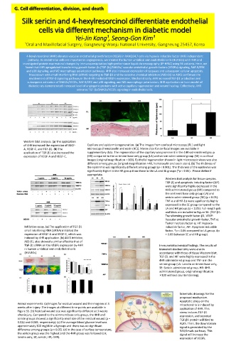
Western blot analysis. (a) The application
of 4HR increased the expression of VEGF- Capillary and epidermis regeneration. (a) The images from confocal microscopy (FL ) and light
A, VEGF-C, and TGF-β1. (b) The microscopy (hematoxylin and eosin (HE)). Movie clips for confocal images are available in
application of TGF-β1 also increased the supplementary data. The regeneration of the capillary was prominent in the 4HR-administered group
expression of VEGF-A and VEGF-C. (HR) compared to the ointment base only group (LA) and sericin-administered group (SE) in confocal
images (original magnification ×100). Epithelial regeneration shown in light microscopic views was also
different among groups (original magnification ×40, hematoxylin and eosin stain). (b) The thickness of
the epidermis was significantly different among groups (p < 0.001). The thickness of the epidermis was
significantly higher in the HR group than those in the LA and SE groups (*p < 0.05). Please define if
appropriate.
Western blot analysis for tissue samples.
TGF-β1 and apoptosis-inducing factor (AIF)
were significantly highly expressed in the
4HR-administered group (HR) compared to
the ointment base only group (LA) and
sericin-administered group (SE) (p < 0.05).
TNF-α and Hif-1α were significantly highly
expressed in the SE group compared to the
LA and HR groups (p < 0.05). Full-length gels
and blots are included in Figure S6. (TGF-β1:
Transforming growth factor-β1, VEGF:
Vascular endothelial growth factor, TNF-α:
Tumor necrosis factor-α, Hif: Hypoxia-
Inhibition assay. (a) The application of TGF-β1 inducible factor, AIF: Apoptosis inducible
small interfering RNA (siRNA) inhibited the factor. *p < 0.05 compared to LA group. #p
expression of VEGF-A and VEGF-C, which was < 0.05 between SE and HR group).
induced by 4HR application. (b) ALK5 inhibitor,
A83-01, also showed a similar effect to that of
TGF-β1 siRNA on the VEGFs expression by 4HR Immunohistochemical findings. The results of
in human umbilical vein endothelial cells immunohistochemistry were also in
(HUVECs). accordance with those of tissue Western blot.
TGF-β1 and AIF were highly expressed in the
4HR-administered group and TNF-α in the
sericin group (LA: Lanolin ointment base only,
SE: Sericin-administered group, HR: 4HR-
administered group, original magnification
×100 without counterstaining).
Schematic drawings for the
proposed mechanism.
Apoptotic stress on the
Animal experiments. (a) Images for residual wound and thermogram at 3 mitochondria is induced by
weeks after injury. The images at different time points are available in
application of 4HR. This
Figure S5. (b) Residual wound size was significantly different at 3 weeks stress induces TGF-β1
after injury. Compared to the ointment base only group, the 4HR and expression, and secreted
sericin groups showed a significantly small size of the residual wound (p = TGF-β1 protein will bind to
0.022 and 0.049, respectively). (c) The average blood glucose level was ALK5. Then, the downstream
approximately 300 mg/dL in all groups and there was no significant signal is generated by the
difference among groups (p > 0.05). (d) In the case of surface temperature, RAS/Smads pathway. This
the sericin group was the highest and the 4HR group was followed (LA; signal will increase the
lanolin only, SE; sericin, HR; 4HR).
expression of VEGFs.