
Page 55 - D. Cancer biology
P. 55
Inhibition of cathepsin D enhances chemosensitivity via RNF183-mediated degradation of
Bcl-xL by autophagosomal IκB degradation
2
1
2
2
Kyoung-jin Min , Seung Un Seo , Seon Min Woo , Taeg Kyu Kwon *
1 New Drug Development Center, Daegu-Gyeongbuk Medical Innovation Foundation (DGMIF), 80 Cheombokro, Dong-gu, Daegu 41061, Korea. Department of
2
Immunology, School of Medicine, Keimyung University, 1095 Dalgubeoldaero, Dalseo-Gu, Daegu 42601, South Korea.
BACKGROUND AIM
Cathepsin D (Cat D) is mainly located in the lysosome, and shows multiple Here, we investigated whether inhibition of Cat D could enhance anti-cancer
biological functions, such as degradation of intracellular and extracellular proteins, drug-mediated apoptosis, and identified the molecular mechanisms that
regulation of cell death, and activation of inflammatory cells. Its role in cancer cells sensitize cancer cells to induce apoptosis by combination treatment involving
is also worth studying, considering its high expression in multiple cancers. Cat D anti-cancer drug and inhibition of Cat D.
increases cancer invasion, metastasis, and angiogenesis, and its overexpression
increases the risk of recurrence and death in female patients with breast cancer.
Although several previous studies had reported the role of Cat D in cancer, its
functions and molecular mechanisms in cancer cell death is not yet clear.
METHODS
Renal carcinoma Caki cells and xenograft mice were treated with anti-cancer drugs in the absence or presence of pepstatin A (Pep A; Cat D inhibitor) to identify the
synergistic effects. siRNA and CRISPR/Cas9-mediated system was used to knockdown/knockout of Cat D in Caki cells to investigate its role as a sensitizer. Western
blotting and reverse transcription PCR was used to detect the expression level of apoptosis-related proteins. Ubiquitination assay performed to investigate the effect
of E3 ligase-dependent modulation of protein expression.
RESULTS
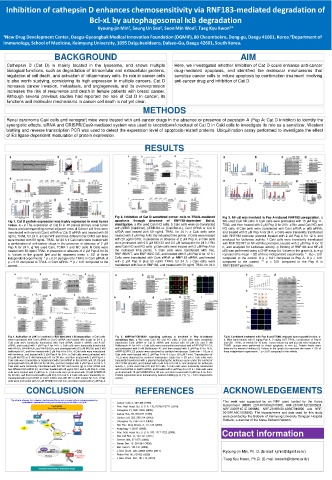
Fig 2. Inhibition of Cat D sensitized cancer cells to TRAIL-mediated Fig 3. NF-κB was involved in Pep A-induced RNF183 upregulation. a
Fig 1. Cat D protein expression was highly expressed in renal tumor apoptosis through decrease of RNF183-dependent Bcl-xL We used Caki KD cells. b Caki cells were pretreated with 15 µM Bay 11-
tissue. a - c The expression of Cat D in 40 paired primary renal tumor stabilization. a We used Caki KO cells. b Caki cells were co-transfected 7082, and then treated with 2 µM Pep A for 24 h. c We used Caki KD and
tissues and corresponding normal adjacent ones. d Cancer cell lines were with pEBB (Caki/Vec), pEBB-Bcl-xL (Caki/Bcl-xL), Cont siRNA or Cat D KO cells. d Caki cells were transfected with Cont siRNA or p65 siRNA,
transfected with control (Cont) siRNA or Cat D siRNA and treated with 50 siRNA and treated with 50 ng/mL TRAIL for 24 h. c Caki cells were and treated with 2 µM Pep A for 24 h. e Cells were transiently transfected
ng/mL TRAIL for 24 h. e Caki WT and three different Cat D KO cell lines treated with 2 µM Pep A for the indicated time points. d Cells were treated with RNF183 promoter plasmid, treated with 2 µM Pep A for 12 h, and
were treated with 50 ng/mL TRAIL for 24 h. f Caki cells were treated with with 20 µg/ml CHX, in presence or absence of 2 µM Pep A. e Caki cells analyzed for luciferase activity. f Caki cells were transiently transfected
a combination of anti-cancer drugs in the presence or absence of 2 µM were pretreated with 0.5 µM MG132 and 2.5 µM lactacystin for 24 h. f We with RNF183 WT or NF-κB Mut promoter, treated with 2 µM Pep A for 12
Pep A for 24 h. g We used Caki, TCMK-1 and MC cells. h Cells were used Caki KD and KO cells. g Caki cells were treated with 2 µM Pep A for h, and analyzed for luciferase activity. g Binding of RNF183 and NF-κB
treated with 50 ng/ml TRAIL in presence or absence of 2 µM Pep A for 24 the indicated time points. h Caki cells were transfected with Vec, p65 was performed using a CHIP assay kit. Values in the graph (a, b, e-g)
h. Values in the graphs (b-f and h) represent mean ± SD of three RNF183/WT, and RNF183/C13S, and treated with 2 µM Pep A for 12 h. i represent the mean ± SD of three independent experiments. * . # p < 0.01
independent experiments. * p < 0.01 compared to TRAIL in Cont siRNA. # Cells were transfected with Cont siRNA or RNF183 siRNA, and treated compared to the control. # p < 0.01 compared to Pep A. # p < 0.01
p < 0.01 compared to TRAIL in Cont siRNA. ** p < 0.01 compared to the with 2 µM Pep A plus 50 ng/ml TRAIL for 24 h. j Caki cells were compared to the control. ** p < 0.01 compared to the Pep A in
control. transfected with Vec or RNF183, and treated with 50 ng/ml TRAIL for 24 h. RNF183/WT promoter.
Fig 4. Activation of JNK is involved in Itch-mediated IκB degradation. a Caki cells Fig 5. AMPK/mTOR/ULK1 signaling pathway is involved in Pep A-induced Fig 6. Combined treatment with Pep A and TRAIL reduced tumor growth in vivo. a-
were transfected with Cont siRNA or Cat D siRNA and treated with drugs for 24 h. b autophagy flux. a We used Caki KD and KO cells. b Caki cells were transiently d, Mice were treated with 5 mg/kg Pep A, 3 mg/kg GST-TRAIL, combination of Pep A
Caki cells were transiently transfected with Cont siRNA, Beclin-1 siRNA, and ATG7 transfected Cont siRNA or Cat D siRNA and treated with 10 µM CQ and 5 nM and GST-TRAIL, or vehicle for 16 days. Tumor volume (a) and size (b) were measured.
siRNA, and treated with 2 µM Pep A for 24 h. c Caki cells were transiently transfected Bafilomycin A1 for 24 h. c Caki cells were transiently transfected with mRFP-EGFP-LC3, TUNEL assays were performed to check apoptosis in vivo (c). Protein levels were
with HA-Ub, HA-K48-Ub, and HA-K63-Ub, and pretreated with 0.5 µM MG132 and 5 nM and pretreated with 5 nM Bafilomycin A1 and then treated with 2 µM Pep A for 18 h. detected by western blotting (d). Values in the graph (a) represent the mean ± SD of
bafilomycin A1, and then treated with 2 µM Pep A for 24 h. d Caki cells were transduced mRFP-EGFP-LC3 puncta were observed by confocal microscope. Scale bar = 10 µm d three independent experiments. * p < 0.01 compared to the vehicle.
with lentivirus, and treated with 2 µM Pep A for 24 h. e Caki cells were pretreated with Caki cells were treated with 2 µM Pep A for 6 h (p.c: 0.5 µM Torin). Translocation of
0.5 µM MG132 or 5 nM bafilomycin A1 for 30 min, and then treated with 2 µM Pep A. f TFEB were observed by confocal microscope. Scale bar = 20 µm e Caki cells were
Caki cells were transiently co-transfected with Cont siRNA or Itch siRNA and HA-Ub and transiently transfected with a plasmid harboring the luciferase gene under the control of
pretreated with 0.5 µM MG132 for 30 min, and then treated with 2 µM Pep A for 24 h. h 4X-CLEAR plasmid, and treated with 2 µM Pep A for 24 h, and analyzed for luciferase
Caki cells were transiently transfected with Itch WT and mutant (g) or Cont siRNA and activity. f, g We used Caki KD and KO cells. h Caki cells were transiently transfected
two different Itch siRNA (h), and then treated with 20 µg/ml CHX and 2 µM Pep A. i Caki with Cont siRNA or AMPK siRNA, and treated with 2 µM Pep A for 24 h. i Caki cells were
cells were treated with 2 µM Pep A. j Caki cells were pretreated with 10 µM SP600125 pretreated with 10 µM SP600125 for 30 min, and then treated with 2 µM Pep A for 24 h.
for 30 min, and then treated with 2 µM Pep A for 24 h. k Caki cells were transiently co- Protein expression were measured by western blotting (a, b, f-i). * p < 0.01 compared to
transfected with Cont siRNA or Cat D siRNA and JNK WT or JNK mutant for 24 h. l Caki control.
cells were pretreated with 10 µM SP60012 for 30 min, and then treated with 2 µM Pep A.
CONCLUSION REFERENCES ACKNOWLEDGEMENTS
The scheme showing the molecular mechanisms that sensitize cancer cells to induce apoptosis by This work was supported by an NRF grant funded by the Korea
combination treatment involving anti-cancer drug and inhibition of Cat D • Cancer Cells 2, 383-388 (1990). Government (MSIP) (2014R1A5A2010008, NRF-2019R1A2C2005921,
• Proc. Natl. Acad. Sci. U. S. A. 115, E2762-E2771 (2018). NRF-2020R1C1C1009889, NRF-2018R1D1A3B07049596 and NRF-
• Oncogene 21, 5951-5955 (2002). 2019R1A2C1008993). The biospecimens and data used for this study
• Cancer Res. 53, 873-877 (1993) were provided by the Biobank of Keimyung University Dongsan Hospital
• Cancer Lett. 323, 208-214 (2012). Biobank, a member of the Korea Biobank Network.
• Oncogene 25, 1967-1973 (2006).
• Nat. Rev. Drug Discov. 8, 111-128 (2009).
• Autophagy 4, 85-87 (2008).
• Proc. Natl. Acad. Sci. U. S. A. 103, 1717-1722 (2006). Contact information
• Nat. Cell Biol. 13, 132-141 (2011).
• Science 306, 271-275 (2004).
• Genes Dev. 13, 284-294 (1999).
• Mol. Cell 21, 135-141 (2006).
• J. Biol. Chem. 286, 22886-22893 (2011). -Kyoung-jin Min, Ph. D. (E-mail: kjmin@dgmif.re.kr)
• Redox Biol. 30, 101422 (2020).
• J. Biol. Chem. 294, 101-115 (2019). -Taeg Kyu Kwon, Ph.D. (E-mail: kwontk@dsmc.or.kr)