
Page 31 - A. Aging
P. 31
TMBIM6 regulates aging-associated hepatic steatosis
Kashi Raj Bhattarai , Hyun-Kyoung Kim , Manoj Chaudhary, Hyung-Ryong Kim , Han-Jung Chae 1*
1
1
2
1 Department of Pharmacology and Institute of New Drug Development, School of Medicine, Jeonbuk National University,
Jeonju, Republic of Korea 54896, College of Dentistry, Dankook University, Cheonan 31116, Republic of Korea
2
Contact: meekasik@gmail.com
Abstract
Aging is a process that involves a very complex mechanism, accompanied by the declining capacity of the cellular machinery, which leads to misfolded and aggregated
proteins, resulting ER Stress. The main objective of our study was to investigate the effect of TMBIM6 (Transmembrane BAX Inhibitor Motif Containing 6) on aging-
associated hepatic steatosis. We categorized wild-type (WT) and TMBIM6 knockout (KO) mice into young, middle, and old groups, and observed the effect on hepatic lipid
accumulation in normal food diet condition. The expression of TMBIM6 mRNA and protein was found to be decreased in aging mice. We observed that the TMBIM6
deficient aged mice gained bodyweight, high hepatic triglyceride level, and increased hepatic lipid droplets when compared with its WT counterparts. TMBIM6 KO aged
mice increased oxidative stress, TUNEL positive cells, expression of cleaved caspases 7 and 12. Loss of TMBIM6 induced aberrant ER stress response following hepatic
cell death. Collectively, these results suggest that aging-associated steatosis is regulated by TMBIM6 through the regulation of ER stress and proteostasis.
Keywords: TMBIM6, aging, steatosis, ER stress
Introduction
ER is an important organelle found in eukaryotic cells that serves various functions, including protein synthesis, protein folding, and transporting of synthesized proteins.
Its physiological perturbations affect its function, increase protein-folding demand, and accumulate unfolded/misfolded proteins inside the ER lumen, which increase ER
stress and trigger the unfolded protein response (UPR) to restore cellular homeostasis. ER stress signals are transduced across the ER membrane by three proximal sensors
of the UPR, inositol requiring element-1 (IRE-1), PKR like ER kinase (PERK) and activating transcription factor 6 (ATF6). All three of these sensors are maintained in an
inactive state at the ER membrane by binding to the ER chaperone BiP (Immunoglobulin binding protein). Aging linked declines in expression and activity of key ER
molecular chaperones and folding enzymes compromise proper protein folding and the adaptive response of the UPR. Aging-associated obesity is one of the pathological
conditions in which we tried to identify the molecular mechanisms involved in it.
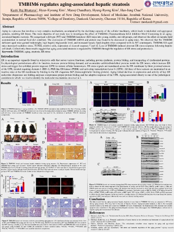
Results Young (3M) Old (20M) A WT TMBIM6 -/- B
A B Young Middle Old Young Middle Old ***
1.5
* 70 ***
60
Actin 1.0 TUNEL 50
40
mRNA/ WT % of TUNEL positive cells 30 **
20
10
TMBIM6 0.5 DAPI 0 Young Middle Old Young Middle Old
-/-
TMBIM6
WT
0.0 Tmbim6 -/- MERGE
Young Middle Old
Figure 1: TMBIM6 declines in aging condition. (A) TMBIM6 mRNA expression was analyzed in the liver of young, middle C WT Liver TMBIM6 - D
and old wild-type mice, (B) TMBIM6 protein level was detected by immunohistochemistry using TMBIM6 antibody in young /- 30 WT 1.2 WT
and old mice. Liver sections of TMBIM6 knock-out mice was used for the validation of antibody. *P<0.05: statistical 42 Y M O Y M O Full length Caspase 12 25 KO 0.9 KO
comparisons. 38 ◀ ◀ Cleaved Caspase 12 20
27 ◀ Cleaved Caspase 12 Cleaved Caspase 12/Actin 15 Cleaved Caspase7/Actin 0.6
A Young Old B WT TMBIM6 -/- 35 ◀ ◀ Full length Caspase 7 10 5 0.3
Cleaved Caspase 7
20
WT TMBIM6 -/- WT TMBIM6 -/- 20x 40x 20x 40x 43 ◀ Actin 0 Young Middle Old 0.0 Young Middle Old
Figure 4: TMBIM6 knock out increase apoptotic response during aging process. (A) TUNEL assay was done to detect
Young apoptotic cells in the liver sections of WT and TMBIM6 KO young and old mice, (B) Bar graph indicates the percentage of
Bhattarai, Kashi Raj, et al. "Endoplasmic Reticulum (ER) Stress Response Failure in Diseases." Trends in Cell Biology 30.9 (2020): 672-675.
TUNEL positive cells, (C) Cleaved caspase 12 and cleaved caspase 7 were detected in WT and TMBIM6 KO liver
Middle staining E homogenates by western blotting, (D) Bar graph indicates the band intensity of figure 3C. *P<0.05, **P<0.01, ***P<0.001
were used for the statistical comparisons.
& WT TMBIM6 -/-
H A B Y M O Y M O
Old Young Old ◀ GRP78
1 2 3 1 2 3
C D ◀ GRP78 ◀ Actin
Young Middle Old Enlarged Young Middle Old Enlarged ◀ CHOP
◀ p-eIF2a
◀ Actin
WT stain WT stain ◀ p-JNK ◀ ATF6a-90
‘O’ Red ◀ Actin ◀ ATF6a-50
TMBIM6 -/- Red Oil TMBIM6 -/- Sirus ◀ CHOP ◀ PERK
◀ p-PERK
◀ Actin
◀ p-eIF2a
◀ eIF2a
Figure 2: TMBIM6 knock out increase hepatic steatosis during aging process. (A) Necroscopic appearance of WT and C ◀ p-IRE1
TMBIM6 KO young and old mice. Yellow arrow indicates abdominal adiposity, (B) Morphological analysis of WT and
TMBIM6 KO liver by H and E staining from different age groups, (C) Oil Red ‘O’ stain was done to demonstrate hepatic lipid WT TMBIM6 -/- ◀ IRE1
◀ sXBP-1
droplets. Yellow arrows indicate hepatic lipid droplets, (D) Sirus Red stain was done to analyze hepatic fibrosis in different age Young Old Young Old
groups of WT and TMBIM6 KO mice. Green arrows indicate the collagen stain. Nucleus sXBP1 ◀ uXBP-1
(55)
A 120 WT **** B Young Middle Old Histone H3 ◀ p-JNK
◀ Bcl-2
****
TMBIM6-/-
◀ PDI
TBARS in liver (micromole MDA/g tissue) 90 * *** *** WT DHE Figure 5: TMBIM6 knock out increase dysregulated ER stress response during aging process. (A) Western blot analysis was
◀ Actin
60
done to detect the ER stress response in the liver lysates of young and old mice using GRP78, p-eIF2 alpha, p-JNK and
30
CHOP. Actin was used as a loading control, (B) Western blot analysis was done to detect the ER stress response in the liver
0
kDa), p-PERK, PERK, p-eIF2 alpha, eIF2 alpha, p-IRE1 alpha, IRE1 alpha, sXBP1, p-JNK, Bcl2, CHOP and PDI were used
Young Middle Old TMBIM6 -/- lysates of young and old mice of WT and TMBIM6 KO genotype. Antibodies such as GRP78, CHOP, ATF6 alpha (90 and 50
C to detect the ER stress response. Actin was used as a loading control, (C) Nuclear fractionation was done from liver lysates,
and spliced XBP1 was analyzed. Histone H3 was used as a nuclear loading marker.
Young Middle Old D Young Middle Old Conclusion
Our data reveals that the aging-associated hepatic steatosis is very high in TMBIM6 KO mice as compared to WT mice
WT during aging process. This may lead to the risk of hepatic fibrosis. Aging-associated steatosis is due to the increased
WT oxidative stress in the liver and is more in TMBIM6 KO mice. Furthermore, the dysregulated UPR response and cellular
8-OHdG 4-HNE apoptosis also contributes to the aging-associated hepatic steatosis. These data suggest that TMBIM6 plays a critical role to
regulate aging and its associated liver disease.
References
Tmbim6 -/- TMBIM6 -/- 1. Bhattarai, Kashi Raj, et al. "Endoplasmic Reticulum (ER) Stress Response Failure in Diseases." Trends in Cell Biology 30.9
(2020): 672-675.
2. Bhattarai, Kashi Raj, et al. "Potential application of ixeris dentata in the prevention and treatment of aging-induced dry
Figure 3: TMBIM6 knock out increase oxidative stress during aging process. (A) Lipid peroxidation was assessed in the liver mouth." Nutrients 10.12 (2018): 1989.
of WT and TMBIM6 KO young and old mice by TBARS assay, (B) ROS was detected in WT and TMBIM6 KO liver sections 3. Naidoo, Nirinjini, and Marishka Brown. "The endoplasmic reticulum stress response in aging and age-related
by DHE staining (10 uM), (C-D) Immunohistochemistry was done in liver sections of WT and TMBIM6 KO mice of various diseases." Frontiers in physiology 3 (2012): 263.
age groups using 8-OHdG (C) and 4-HNE (D) antibodies to detect oxidative stress. *P<0.05, **P<0.01, ***P<0.001 and 4. Salminen, Antero, and Kai Kaarniranta. "ER stress and hormetic regulation of the aging process." Ageing research
*P<0.05, **P<0.01, ****P<0.0001 were used for statistical comparisons.
reviews 9.3 (2010): 211-217.