
Page 27 - A. Aging
P. 27
A-16
Identification of HPOB as a potent inhibitor of tyrosinase
)
)
),
Hyerim Song¹ , Yun Jeong Hwang¹ , Jae Won Ha¹ , Yong Chool Boo¹ *
)
Department of Molecular Medicine, Cell and Matrix Research Institute, BK21 Plus KNU Biomedical Convergence Program,
School of Medicine, Kyungpook National University, Daegu, Republic of Korea
BACKGROUND RESULTS
Dysfunctions associated with melanin synthesis cause clinically relevant
pigmentation disorders that can be congenital or acquired, cutaneous or
systemic, temporary or permanent, and related to hypo- or
hyperpigmentation
Tyrosinase (TYR) is a copper-containing enzyme that catalyzes the
hydroxylation of L-tyrosine to L-3,4-dihydroxyphenylalanine (DOPA), and the
oxidation of L-DOPA to its corresponding o-quinone derivative in the melanin
synthetic pathway. A variety of natural and synthetic compounds are known to
inhibit the catalytic activity of TYR by multiple mechanisms. For example, β-
arbutin and kojic acid are known to inhibit the catalytic activity of TYR in a
competitive manner TYR.
AIM
The present study was undertaken to screen an epigenetic drug library to
identify new drug candidates for pharmacotherapy of hyperpigmentation.
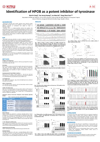
Of 141 total drugs tested in this study, K8 (4-((hydroxyamino)carbonyl)-N-(2- Fig. 6. Effects of K8, b -arbutin and kojic acid on the catalytic activity of
hydroxyethyl)-N-phenyl-benzeneacetamide; HPOB) was found to inhibit α- Fig. 1. Effects of drugs on melanin synthesis and viability in B16-F10 cells. human tyrosinase (TYR) in vitro.
MSH-induced melanogenesis in B16-F10 murine melanoma cells at 1.0 μM Open and closed histograms represent the absence and presence of α-
without significant cytotoxicity. K8 is known to inhibit histone deacetylase melanocyte-stimulating hormone (α-MSH), respectively. Drugs that inhibited
(HDAC) 6 through zinc chelatingactivity. melanin synthesis without cytotoxicity how are highlighted with red color.
We initially expected K8 to be able to reduce melanin by inhibiting TYR
expression by epigenetic mechanisms, but unexpectedly, no strong evidence
for this action could be found. On the other hand, K8 was shown to strongly
inhibit the catalytic activity of TYR in a competitive manner, probably through
copper chelatingactivity.
Here we report that screening of an epigenetic drug library resulted in the
identification of new drug candidates for the pharmacotherapy of skin
hyperpigmentation disorders.
METHODS Fig. 7. Enzyme kinetics for the inhibition of TYR activity by K8 and b-arbutin.
Assays were also performed in the presence of K8 (a) or b-arbutin (b) at the
Reagents and a Drug Library indicated concentrations (μM). Lineweaver-Burk plots are drawn using the
An epigenetic screening library (Item No. 11076) was purchased from Cayman mean values of three different measurements in order to determine the
Chemical (Ann Arbor, MI, USA).
inhibition types of K8 and b-arbutin.
Cell Culture
B16-F10 murine melanoma cells (ATCC CRL-6475) obtained from the American
Type Culture Collection (Manassas, VA, USA) were cultured in Dulbecco’s Fig. 2. Effects of H4 and K8 on the melanin synthesis and viability of B16-F10
modified Eagle’s medium (DMEM) containing 10% fetal bovine serum and cells.
antibiotics (100 U·mL−1 penicillin, 0.1 mg·mL−1 streptomycin, 0.25 μg·mL−1
amphotericin B).
Screening Assay for Overall Melanin Synthesis
B16-F10 cells were plated in 96-well plates (3 × 10³ cells per well) and
cultured for 24 h. The cells were then treated with a test drug at 1.0 μM for 60
min, and subsequently stimulated with 0.1 μM α-MSH for 72 h.
Cell Viability Assay
Cell viability was evaluated using 3-(4,5-dimethylthiazol-2-yl)-2,5-diphenyl Fig. 8. Comparison of copper chelating activities of K8, kojic acid and b-
tetrazolium bromide (MTT). arbutin. (a) Absorption spectra of 200 μM pyrocatechol violet in the absence
and presence of 200 μM CuSO4 and other additives (200 μM). (b)
Melanin Content Assay Pyrocatechol violet (200 μM) was reacted with 200 μM CuSO4 in the presence
The intracellular melanin retained in cells and extracellular melanin secreted of varied concentrations of K8, kojic acid and b-arbutin.
from cells were separately determined. Fig. 3. K8 reduces the melanin content and tyrosinase (TYR) activity in B16-
F10 cells stimulated with α-MSH.
Cellular TYR Activity Assay
After B16-F10 cells were treated with drug and/or α-MSH, the TYR activity of
cell extracts was determined by an indirect spectrophotometric method to
monitor dopachrome formation with L-tyrosine plus L-3,4-dihydroxyphenyl
alanine (L-DOPA) as the substrates.
Quantitative Reverse Transcription Polymerase Chain Reaction (qRT-PCR)
Analysis
Table 1. Sequences of primers used for qRT-PCR.
Gene GenBank Accession
Name Number Primer Sequences
Forward: 5ʹ-CTTCTTCTCCTCCTGGCAGATC-3ʹ
TYR NM_011661.5
Reverse: 5ʹ-TGGGGGTTTTGGCTTTGTC-3ʹ
Forward: 5ʹ-CAGTGCAGCGTCTTCCTGAG-3ʹ
TYRP1 NM_001282015.1
Reverse: 5ʹ-TTCCCGTGGGAGCACTGTAA-3ʹ
Forward: 5ʹ-GCAAGAGATACACGGAGGAAG-3ʹ
DCT NM_010024.3
Reverse: 5ʹ-CTAAGGCATCATCATCATCACTAC-3ʹ
Forward: 5ʹ-GCTGGAAATGCTAGAATACAG-3ʹ Fig. 9. Effects of drugs on human TYR activity in vitro. The activity of human
MITF NM_008601.3 TYR was determined in the absence or presence of each drug (1.0 µM). Drugs
Reverse: 5ʹ-TTCCAGGCTGATGATGTCATC-3ʹ
Forward: 5ʹ-GCATCTCCCTCACAATTTCCA-3ʹ that inhibited the catalytic activity of TYR in vitro are highlighted with red
GAPDH NM_001289726.1
Reverse: 5ʹ-GTGCAGCGAACTTTATTGATGG-3ʹ color.
Western Blotting CONCLUSION
Western blotting was performed as previously described [26,35]. Primary Fig. 4. Effects of K8 on the mRNA and protein levels of TYR, tyrosinase-related In conclusion, the present study identified K8 from an epigenetic drug library
antibodies for TYR (#127217) was purchased from MyBioSource (San Diego, protein 1 (TYRP1) and dopachrome tautomerase (DCT) in B16-F10 cells as a potent inhibitor of cellular melanin synthesis, suggesting its potential
CA, USA). Primary antibodies for TYRP1 (#10443) and β-actin (#47778) were stimulated with α-MSH. utility in the treatment of hyperpigmentation disorders. The results suggest
purchased from Santa Cruz Biotechnology (Santa Cruz, CA, USA). Primary that K8 inhibits cellular melanin synthesis not through “epigenetic” down-
antibodies for MITF (#20663) and DCT (#74073) were purchased from Abcam regulation of TYR protein levels but by direct inhibition of TYR catalytic activity.
(Cambridge, UK).
Further studies are needed to examine its in vivo and clinical efficacy.
In vitro TYR Catalytic Activity Assay
Lysate of B16F10 cells stimulated with α-MSH for 24 h was used as a murine REFERENCES
TYR preparation. Human TYR preparation was the lysate of human embryonic 1. Kim JH, Seok JK, Kim YM, Boo YC. Identification of small peptides and
kidney 293 cells constitutively expressing human TYR (HEK293-TYR. glycinamide that inhibit melanin synthesis using a positional scanning
synthetic peptide combinatorial library. British Journal of Dermatology.
Copper Chelating Activity Assay 2019l;181:128-137.
Copper chelating activity was determined by using pyrocatechol violet. 2. Kwak JY, Seok JK, Suh HJ et al. Antimelanogenic effects of luteolin 7-sulfate
isolated from Phyllospadix iwatensis Makino. British Journal of
Statistical Analyses Dermatology 2016; 175: 501-11.
The data are expressed as the mean ± standard deviation (SD) of three
independent experiments. The experimental results were statistically analyzed ACKNOWLEDGEMENT
using SigmaStat v. 3.11 software (Systat Software Inc., San Jose, CA, USA), by Fig. 5. Effects of K8 on the mRNA and protein levels of microphthalmia- This work was supported by a National Research Foundation of Korea (NRF)
one-way analysis of variance (ANOVA), followed by Dunnett’s test. associated transcription factor (MITF) in B16-F10 cells stimulated with α-MSH. grant funded by the Korea government (Ministry of Science and ICT, No.
2019R1I1A2A01045132).