


Page 289 - ebook
P. 289
F-1
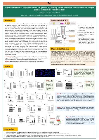
Nephronophthisis 3 regulates cancer cell growth by primary cilium formation through reactive oxygen
species-induced HIF-1alpha and Erk
Jae-Wook Lee and Eun-Yi Moon
Department of Bioscience and Biotechnology, Sejong University, Seoul, 05006, Republic of Korea
Abstract Nephrocystin-3 (NPHP3)
Primary cilium is antenna-like organelle projecting from the surface of cell membrane.
We reported previously that primary cilium formation could be regulated by • Mutations in the mouse ortholog
nephronophthisis 3 (NPHP3) expression followed by its interaction with by thymosin β4 of human NPHP3 cause poly
using HeLa cervical cancer cells. Here we investigated whether cancer cell growth could cystic kidney disease (pcy) in
be regulated by NPHP3 expression-associated primary cilium formation. Cancer cell mouse model
growth was retarded by the incubation without fetal bovine serum (FBS). The number of • Disruption of NPHP3 alters the
cells with primary cilia was increased by serum starvation which enhanced NPHP3 frequency and/or the length of
expression. While NPHP3 expression was inhibited by siHIF-1α, it was increased by the primary cilia
incubation of cells under hypoxic condition. Under serum starvation, as reactive oxygen
species were elevated, the number of cells with primary cilia and an increase in NPHP3 • Loss of Nephrocystin-3 function
expression were inhibited by N-acetylcysteine treatment. The number of cells with can cause Embryonic Lethality,
primary cilia was increased by H 2 O 2 treatment, which was inhibited by siHIF-1α. The Meckel-Gruber-like and Syndrome,
Inversus,
Situs
Renal-
number of cells with primary cilia and NPHP3 expression were decreased by the Hepatic-Pancreatic Dysplasia
inhibition of Erk with P98059. Cancer cell growth was reduced by the treatment with Figure 1. Allergic responses in mast cells..
cilliobrevin A. Taken together, the results imply that the number of cancer cells with
primary cilia might be controlled by NPHP3 expression under serum starvation. Data also Methods & Materials
demonstrate that NPHP3 expression could be regulated by ROS-induced HIF-1 and
Erk in HeLa human cervical cancer cells. It suggests that cancer cell growth under serum Detection of primary cilia was measured as follow. HeLa cells were grown on coverslip and
starvation could be associated with the regulation of NPHP3 expression to maintain then incubated with serum-starved DMEM with 0.1% FBS for 36 h. Cells were fixed with 4%
paraformaldehyde for 10 min, washed three times with cold PBS, and permeabilized with
primary cilium formation. PBST (0.1% (v/v) Triton X-100 in PBS) for 10 min. Then, cells were washed three times, and
incubated with monoclonal anti-acetylated tubulin antibodies diluted (1:1000) in PBST for 1
Acknowledgement: This work was supported by grants from the Basic Research Laboratory h at room temperature. After washing three times with PBS, cells were incubated with FITC-
Program (#2021R1A4A5033289) through the National Research Foundation of Korea (NRF) conjugated goat anti-mouse IgG-secondary antibody or goat anti-mouse IgG-Alexa 568
funded by the Ministry of Education, Science and Technology (MEST), Korea. diluted (1:1000) in PBST for 1h at room temperature. Nucleus was visualized by staining cells
with DAPI. After washing with PBS, cells were mounted on glass slide. Primary cilia were
Keywords: Nephronophthisis 3, Primary cilium, HIF-1, Erk, Reactive oxygen species observed and photographed at 1000 x magnification under a fluorescence microscope
(Nikon, Tokyo, Japan).
Cytotoxicity was measured by luminescence assay.using Celltiter-glo substrate.
Results
A B C D Figure 2. Total cell number was reduced
by serum starvation (A) but no changes
in cell viability (B). Ciliogenesis was
increased by serum starvation (C).
NPHP3 expression was enhanced by
serum starvation (D).
A C D E Figure 3. HIF- A B C
B 1 increased Figure 4. HIF-1
by serum under hypoxic
starvation (B) condition (A-B)
or DMOG (D) contribute to
contribute to ciliogenesis (C).
ciliogenesis (A,
C, E).
A B C D E
Figure 5. Reactive oxygen species (ROS) enhanced ciliogenesis. ROS was
increased under serm starvation (A). ROS-mediated cilliogenesis was reduced
by N-acetylcysteine (NAC) (B). NAC also inhibited ciliogenesis-associated
NPHP3 (C). ROS-mediated cilliogenesis was confirmed by the treatment with
hydrogen peroxide (H 2 O 2 ) (D). Cilliogenesis by H 2 O 2 treatment was mediated
by HIF-1, which was confirmed by the treatment with HIF-1 -siRNA (E).
A C D
B
Figure 6. Serum starvation increased Erk phosphorylation (A), which was
inhibited by NAC treatment (B). PD98059, Erk inhibitor, reduced NPHP3
transcriptional activity (C) and ciliogenesis (D)..
A B C D D
Figure 7. Cilliobrevin A, inhibitor for primary cilium
formation, inhibited ciliogenesis (A), NPHP3 expression
and transcriptional activity (B), and cell viability (C). This
scheme demonstrates that NPHP3 expression could be
regulated by ROS-induced HIF-1 and Erk which lead to
the control of cancer cell viability (D)
Summaries
1. The number of cancer cells with primary cilia might be controlled by NPHP3 expression under serum starvation.
2. Data demonstrate that NPHP3 expression could be regulated by ROS-induced HIF-1 and Erk in HeLa human cervical cancer cells.
3. It suggests that cancer cell growth under serum starvation could be associated with the regulation of NPHP3 expression to maintain primary cilium formation.
4. Therefore, it is required the comprehensive studies on the regulation of cancer cell viability.

