


Page 36 - ebook
P. 36
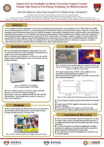
Impact of X-ray Irradiation on Blood Cells using Compact Ceramic
                   Vacuum Tube based on Fast Pulsing Technology for Blood Irradiator


                     Jinho Choi¹, Hanna Lee², Jaekyu Jang³, Kyung-Sik Yoon⁴, Moonkyoo Kong⁵*, Jehwang Ryu⁶*
                                1 Department  of Radiation  Oncology,  College of Medicine, Kyung  Hee University,  Seoul 02447, Korea;
                             2 Department  of Age Service-Tech  Convergence,  College of Medicine, Kyung  Hee University,  Seoul 02447, Korea;
            3 R&D, CAT Beam Tech Co., Seoul 02455, Korea; 4 Department  of Biochemistry  and Molecular  Biology,  College of Medicine, Kyung  Hee University,  Seoul 02447, Korea;
       5 Department  of Radiation  Oncology,  Kyung  Hee University  Medical Center, Seoul 02447, Korea; 6 Department  of Radiology,  College of Medicine, Kyung  Hee University,Seoul,02447,  Korea


               Abstract

       X-ray irradiation of blood components is used to prevent transfusion-associated graft-versus-host disease (TA-GVHD). When TA-GVHD
      occurs, clinical symptoms such as fever, rash, pancytopenia, liver dysfunction, edema, and systemic blisters appear within 10 to 12 days after
      transfusion. Once the disease develops, there is no effective treatment, so prevention is essential. In order to prevent graft-versus-host disease
      caused by blood transfusion, X-ray irradiation is performed on blood products. Medical institutions have facilities with blood irradiators for
      the irradiation of blood components and use a dose of 20 to 50 Gy. In this study, we developed a compact vacuum X-ray tube sealed by the
      ceramic body instead of a glass envelope. Piranha Multimeter was used to measure kVp, Time, HVL, Total Filtration, Dose, and Dose rate.
      After the characteristics of the X-ray tube were obtained, the X-ray was irradiated to blood cells. In this study, we propose a method to
      reduce unnecessary doses to medical staff by irradiating X-rays with a digital radiation source.

              Introduction                                                  Results


      ◈ X-rays have high penetrating power like gamma rays, and
      like electron beams, they can be controlled by turning the power
      on/off, so they can be used easily and safely.
      ◈ The conversion energy efficiency was very low, so it was still
      mainly used for medical diagnosis, but it was overcome with fast
      pulsing technology.

         (A)                    (B)
                                                                 Fig 3. (A) Manufactured monoblock filled with insulating oil for the shot
                                                                 test, (B) The moment of X-ray exposure with X-ray tube in test bed when
                                                                             shot test is conducted in high voltage.
                                                                 ◈ To apply a high voltage of 70kV to the monoblock, it was
                                                                 carried into insulating oil shown in Figure 3 (A).
                                                                 ◈ The performance evaluation of the X-ray tube was measured
                                                                 with a Piranha multimeter.


                  Fig 1. (A) RADGIL2 X-ray irradiator,
                 (B) X-ray blood irradiator X-RAD 3000.
      ◈ X-ray irradiator system is an effective, modern and safer
      alternative to Cesium-137 sourced irradiators.
      ◈ Several kV values are available from 80, 100, 120, 140, 160,
      180 and 200 kV. Also, Possible tube current, mA values, are   Fig 4. (A) Tube voltage and (B) Exposure rate of ceramic x-ray tube
      from 5, 10, 15 and20 mA.
                                                                ◈ 1 Cycle consists of exposure time 2s, interval time 18s and total
                                                                20s.
                 Methods                                        ◈ A high voltage of 70 kV applied to the anode was successfully
                                                                driven for 300 times.
                                                                ◈ It was confirmed that the compact ceramic X-ray tube
      ◈ The envelope material is different. One is an X-ray tube with a
      glass envelope, and the other is an X-ray tube with a ceramic   irradiated the dose without significant fluctuation for 300 cycles.
      envelope manufactured by CAT Beam Tech.
                                                                          Conclusion & Discussion

                                                                ◈ There are several advantages when the envelope of X-ray tube
                                                                is ceramic. Using ceramic-enveloped tubes instead of glass for X-
                                                                ray applications will provide better durability and reduce the size
                                                                and volume.
                                                                ◈ We have made efforts to develop the application by making
                                                                monoblock by applying an insulating material that can perform
                                                                all the functions of insulating oil and replaceable.
                                                                ◈ We conducted as a basis for developing an X-ray tube for use
        Fig 2. Vacuum sealed X-ray tubes of to compare the size of the tubes  in a blood irradiator.

