


Page 21 - ebook
P. 21
DNA-PKcs regulates immune signaling
triggered by mitochondrial DNA double-stranded Breaks
June Heo 1,2 Ho-Soo Lee and Hyeseong Cho 1,2
1
1 Department of Biochemistry and Molecular Biology, Ajou University School of Medicine, Suwon, South Korea
2 Department of Biomedical Sciences, Graduate School of Ajou University, Suwon, South Korea
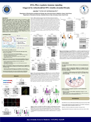
ABSTRACT Figure 1. Mitochondrial transcription factors with FokI endonuclease decrease a
copy number of mitochondrial DNA. a, Mitochondrial DNA-targeted FokI endonucleases
Mitochondrial DNA (mtDNA) encodes necessary genes for the generation of ATPs
(adenosine triphosphates), which are used as an energy source for intracellular (mito-FokI) are fusion proteins composed of mitochondrial transcription factors, FokI cleavage
activities. mtDNA is prone to double-stranded breaks damage caused by replication domain, and mCherry fluorescent protein. b, Western blot analysis using HeLa cell line to
errors, ultraviolet light, radiation, and various chemicals. Cleaved mtDNA is rapidly confirm the expression of mito-FokI. WT, wild type; MT, mutant, catalytic dead mutant of FokI
degraded by mitochondrial replication machinery. During the processing of c, Western blot analysis after subcellular fractionation for localization of mito-FokI. N, nuclear
mitochondrial DNA double-strand breaks (mtDSBs), some of mitochondrial nucleic acids fraction; C, cytosolic fraction; M, mitochondrial fraction. d, Immunocytochemistry for mito-FokI
are released into the cytoplasm and then trigger an aberrant innate immune response. in HeLa cell lines stably expressing mito-pDendra-2 fluorescent protein. e, Immunocyto-
Persistency of immune signaling leads to host immunopathology. Here, using
mitochondrial DNA-targeted FokI endonucleases, we showed that mtDSBs triggered chemistry of mito-FokI with antiDNA antibody. f, Western blot analysis of endogenous TFAM
mtDNA degradation and activated type-I interferon response and interferon-stimulated protein in HEK293T cell line. g, Quantitative PCR to confirm relative mtDNA copy numbers
genes (ISGs) through DNA-PKcs-HSPA8 signaling axis. We also explored protein H for each sample using primer in a sequence of D-Loop, MTCOX2. Normalized expression
regulating mtDSBs-induced immune signaling and showed that reduced expression of data are mean ± SEM. derived from n = 4 technical replicates. Statistics was performed
mRNA of interferon β1 and ISGs in response to overexpressing protein H. Our data using One-way ANOVA with confidence interval = 95%; *P ≤ 0.05, **P ≤ 0.01, ***P ≤ 0.005,
suggest that protein H acts as a key molecule regulating mtDSBs-induced immune ns, nonspecific.
signaling dependent on the DNA-PKcs-HSPA8 axis.
INTRODUCTION
mtDNA stress
mtDNA degradation and
release of mitochondrial nucleic acids
Activation of cytoplasmic sensors
➢ mtDNA (cGAS–STING)
➢ mtRNA (RIG-I–MDA-5–MAVS)
Activation of interferon (IFN) regulatory factors
Secretion of Type I interferon
Innate immunity upon mtDNA dysfunction
Figure 4. Protein H degrade activated DNA-PKcs in a proteasome-dependent manner for
▪ Various sources of mtDNA stress including mitochondrial DSBs trigger mtDNA suppression of type-I IFN signaling. a, Western blot analysis of pHSPA8 in HEK293T cells
dysfunction and degradation. co-transfected with TFAM-FokI WT and Protein H in a dose-dependent manners and catalytic
dead mutant. b, Western blot analysis of pDNA-PKcs in HEK293T cells co-transfected TFAM-
- Mitochondrial DNA stress primes the antiviral innate (2015) Nature FokI WT and catalytic active mutant of Protein H. c, RT-qPCR of IFNB1, RSAD2 in HEK293T
- The mitochondrial DNA polymerase gamma degrades linear DNA fragments precluding cells co-transfected with TFAM-FokI and Protein H; d, co-transfected with TFAM-FokI and
the formation of deletions (2018) Nat. Commun. Protein H WT or Protein H active or Protein H dead mutant. Normalized expression data are
- Linear mitochondrial DNA is rapidly degraded by components of the replication mean ± SEM. derived from n = 4 technical replicates. e, West blot analysis of pDNA-PKcs and
machinery (2018) Nat. Commun.
pHSPA8 in HEK293T cells treated with a proteasome inhibitor(MG132) or autophagy
▪ BAK/BAX pores, VDAC oligomers and the mitochondrial permeability transition pore inhibitor(bafilomycin) in addition to co-transfected with TFAM-FokI and Protein H.
(mPTP) serve as channel for the release of mitochondrial nucleic into the cytoplasm.
- BAK,BAX macropores facilitate mitochondrial herniation and mtDNA efflux
during apoptosis (2018) Science
- VDAC oligomers form mitochondrial pores to release mtDNA fragments and promote CONCLUSION
lupus-like disease (2019) Science
The cleaved mitochondrial DNAs by mito-FokI are predominantly
▪ Similarly, stress imposed on the RNA granules (MRG) by perturbation of mitochondrial
dynamics or deletion of RNA processing enzymes drives the escape of dsRNA from the degraded.
mitochondrial matrix.
Howevers, some of fragmented mtDNAs are released into the
- Mitochondrial double-stranded RNA triggers antiviral signalling in humans (2018) Nature cytoplasm through the oligomeric VDAC pore and then activate
- Nuclear sensing of breaks in mitochondrial DNA enhances immune surveillance (2021) Nature
an immune response.
• Released mitochondrial nucleic acids activate cytosolic sensors and then innate
immune response expressing type-I interferon and proinflammatory cytokines. • The cytoplasmic mtDNA fragments activate DNA-PKcs-HSPA8 axis immune
Figure 2. FokI endonucleases targeting the mtDNA induce the activation of DNA-PKcs- response and this event culminates in the release of type I interferon and
HSPA8 axis pathway through the VDAC pore protein. a, Western blot analysis of pIRF3 paracrine signaling.
and pHSPA8 in HEK293T cells transfected with MTERF1-FokI WT or catalytic MT b, TFAM-
RESULTS FokI WT or catalytic MT in the indicated a time-dependent manner. c, West blot analysis of
pDNA-PKcs(S2056), pHSPA8, and pIRF3(S386) in cells transfected with TFAM-FokI WT;
d, treated with DNA-PKcs inhibitor(Nu-7026) in c. e, Timeline of the mtDSBs in response to
knockdown of VDAC pore protein. f, Western blot analysis to confirm the knockdown of VDAC
(left), and expression of pHSPA8 in response to TFAM-FokI WT or MT with knockdown of
VDAC (right); g, or treated with an inhibitor of VDAC oligomerization.
Protein H serves as negative regulator of cGAS-independent
immune signaling.
• DNA-PKcs sensing cytosolic mtDNA is degraded by Protein H in a dose-
dependent manner.
• mtDSBs-induced immune response including mRNA of IFNB1 and interferon
stimulated genes(ISGs) is downregulated by Protein H.
REFERENCES
- Human DNA-PK activates a STING-independent DNA sensing pathway (Sci Immunol. 2020)
- Cytoplasmic DNA sensing by KU complex in aged CD4+ T cell potentiates T cell activation
and aging-related autoimmune inflammation (Immunol. 2021)
Figure 3. Activation of the DNA-PKcs-HSPA8 axis triggers a cGAS-independent immune
response. a, Western blot analysis to confirm a knockout(KO) of cGAS in HeLa cell line. b, RT- - Mechanisms of Mitochondrial DNA deletion formation (Trends genet. 2019)
qPCR of IFNB1 in cGAS KO HeLa cells transfected with MTERF1-FokI or TFAM-FokI in the
indicated a time-dependent manner. c, RT-qPCR of IFNB1, IFI44 and IFIT1 in cGAS KO HeLa - Safeguarding mitochondrial genomes in higher eukaryotes (Nat. Struct. Mol. Biol. 2020)
cells treated with DNA-PKcs inhibitor(Nu7026) in addition to transfected with MTERF1-FokI WT
or TFAM-FokI WT. Normalized expression data are mean ± SEM. derived from n = 4 technical
replicates.

