


Page 15 - ebook
P. 15
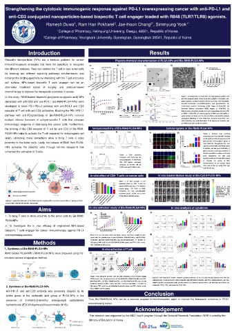
Strengthening the cytotoxic immunogenic response against PD-L1 overexpressing cancer with anti-PD-L1 and
Poster Title
anti-CD3 conjugated nanoparticles-based bispecific T cell engager loaded with R848 (TLR7/TLR8) agonists.
Researchers’/Presenters’ Names
Ramesh Duwa , Ram Hari Pokharel , Jae-Hoon Chang , Simmyung Yook 1*
1
2*
2
Institution/Organization/Company
1 College of Pharmacy, Keimyung University, Daegu, 42601, Republic of Korea
2 College of Pharmacy, Yeungnam University, Gyeongsan, Gyeongbuk 38541, Republic of Korea
Introduction Results
Bispecific nanoparticles (NPs) are a modular platform for cancer Physico-chemical characterization of PLGA-NPs and Bis-R848-PLGA-NPs
immunotherapeutic strategies that have the specificity to recognize A) B) C) D) E)
two different epitopes. They can redirect the T cell to lyse tumor cells 200 Particles sizes 0.4 -15 Loading capacity (%)
PDI
Encapsulation efficiency (%)
by blocking two different signaling pathways simultaneously and 150 0.3 Poly-dispersity index (PDI) Zeta potential (mV) -20 10 60
8
0.2
100
40
enhance the binding specificity by interacting with the T cell and tumor Particles size (nm) 50 0.1 -25 Loading capacity (%) 6 20 Encapsulation efficiency(%)
4
cell surface. NPs-based bispecific T cells engager can be an 0 0.0 -30 2 L L L L L L 0
Bis-R848-PLGA-NPs
R848-PLGA-NPs
Anti-PD-L1-R848-PLGA-NPs
R848-PLGA-NPs
Bis-R848-PLGA-NPs
Anti-PD-L1-R848-PLGA-NPs
0
50
100
200
alternative treatment option of surgery and platinum-based Free PLGA-NPs Anti-CD3-R848-PLGA-NPs Free PLGA-NPs Anti-CD3-R848-PLGA-NPs 10 20 Concentration of R848 500
chemotherapy to improve the therapeutic outcomes in cancer.
F) G) H)
In this study, R848-loaded bispecific poly(lactic-co-glycolic acid) NPs 100 300 Figure 1. Characterization of PLGA-NPs. (A) Hydrodynamic particle size
and Poly-dispersity index of free PLGA-NPs, anti-PD-L1-PLGA-NPs, anti-
decorated with anti-CD3 and anti-PD-L1 (bis-R848-PLGA-NPs) were 80 250 CD3-PLGA-NPs, and Bis-PLGA-NPs. (B) Zeta potential of free PLGA-NPs,
200
60
anti-CD3-PLGA-NPs,
anti-PD-L1-PLGA-NPs,
(C)
and
Bis-PLGA-NPs.
developed to block PD-1/PD-L1 pathway with anti-PD-L1 and CD3 Cummulative release % 40 Acidic pH (5.4) Mass of conjugated Abs on PLGA-NPs (g/mg) 150 Transmission electron microscopy (TEM) images of PLGA-NPs. (D)
100
receptor of T cell with anti-CD3 antibodies. Blocking the PD-1/PD-L1 20 Basic pH (7.4) 50 Scanning electron microscopy (SEM) images of PLGA-NPs. (E)
0
Determination of Loading capacity (LC) and encapsulation efficiency (EE)
0
0 24 48 72 96 120 144 168 192 216 100 200 250 300 500 of R848 in PLGA-NPs. (F) Release profile of R848 from PLGA-NPs in
Concentration of Abs (g/mL)
pathway with anti-PD-L1antibody of Bis-R848-PLGA-NPs restores Time(hrs) Free PLGA-NPs Anti-PD-L1-PLGA-NPs acidic pH(5.4) and basic pH (7.4). (G) Anti-PD-L1 and anti-CD3 antibody
Anti-CD3-PLGA-NPs Bis-PLGA-NPs
multiple effector functions of antigen-specific T cells that enhance conjugation efficiency on free PLGA-NPs, anti-PD-L1-PLGA-NPs, anti-
CD3-PLGA-NPs, and Bis-PLGA-NPs(H) FT-IR spectra of PLGA-NPs and
immunologic response in destroying the cancer cells. Furthermore, anti-PD-L1, anti-CD3, and Bis-PLGA-NPs.
the binding of the CD3 receptor of T cell by anti-CD3 of Bis-R848- Immunoreactivity of Bis-R848-PLGA-NPs Cellular uptake of Bis-R848-PLGA-NPs
PLGA-NPs helps to activate the T cell response for immunogenic cell A) SK-OV3 B) B16F10 A) B) Figure 3. Confocal laser scanning
microscopy and flow cytometry analysis
death. Ultimately, these interactions allow to bring T cells in close for the qualitative (A) and quantitative (B)
SK-OV3 SK-OV3 determination of intracellular uptake of
proximity to the tumor cells. Lastly, the release of R848 from PLGA- free PLGA-NPs, Cou-6-PLGA-NPs, anti-
NPs activates the dendritic cells through toll-like receptor-8 that CD3-Cou-6-PLGA-NPs, anti-PD-L1-Cou-6-
PLGA-NPs, and Bis-Cou-6-PLGA-NPs, (1
enhanced the activation of T cells. C) µl/mL equivalent cou-6 concentration) on
CD8 T cell Figure 2 Flow cytometry SK-OV3 and B16F10 cell lines at 6 h time
+
histogram plot illustrating the B16F10 point respectively. The cells were
incubated with Cou-6-loaded NPs (green)
immunoreactivity of Bis-PLGA- B16F10
to visualize the uptake of NPs.
NPs (250 µm/mL equivalent
Counterstaining of cell nucleus (blue)
antibody) (A) SK-OV3 cell (B)
color was carried out using DAPI dye;
B16F10 cell and (C) CD8 T cell.
+
scale bar (20 μm).
In vitro effect of CD8 T cells on cancer cells In vivo biodistribution study of Bis-Cy5.5-PLGA-NPs
+
120 Max
Figure 4. In vitro analysis of cell
100 *** cytotoxity effect of CD8 + T cells on
Cell viability % 60
&
Cell 80 cancer (B16F10 cell line. (*** P<0.001 vs Figure 5. In vivo
death 40 $ control group. ᴨᴨᴨ P< 0.001 vs R848- biodistribution study
the
showing
high
nanoBiTE (Bis-R848-PLGA-NPs 20 PLGA-NPs, & P< 0.01 anti-CD3-R848- penetration and
$
0 PLGA-NPs group, and P< 0.01 vs anti- accumulation of bis-
Cy5.5-PLGA-NPs
in
Resiquimod (R848) Control Free PLGA-NPs PD-L1-R848-PLGA-NPs group). tumor tissue.
R848-PLGA-NPs
Anti-PD-L1-R848-PLGA-NPs
Scheme 1. Systematic illustration of Bis-R848-PLGA-NPs targeting PD-L1 on tumor cell and CD3 on T cell by anti-PD-L1 B16F10 + Tcell+ DC cell Anti-CD3-R848-PLGA-NPs Bis-R848-PLGA-NPs
and anti-CD3 of Bis-R848-PLGA-NPs, respectively. Min
Aims In vivo antitumor study of Bis-R848-PLGA-NPs In vivo analysis of cytokines
A) PBS B) A) B)
Free PLGA-NPs 16
1. To bring T cells in close proximity to the tumor cells by Bis-R848- 8000 R848-PLGA-NPs 14
Anti-CD3-R848-PLGA-NPs
12
Anti-PD-L1-R848-PLGA-NPs 10 ###
Bis-R848-PLGA-NPs
6000
$$$
PLGA-NPs Tumor volume (mm 3 ) 4000 Tumor weight (g) 8 ***
6
4
2
2. To investigate the in vivo efficacy of engineered NPs-based 2000 0 PBS Free PLGA
R848-PLGA-NPs
Anti-PD-L1-R848-PLGA-NPs
bispecific T cells engager for cancer immunotherapy against PD-L1 0 0 2 Times after frist treatment (days) 18 20 Anti-CD3-R848-PLGA-NPs Bis-R848-PLGA-NPs
12
14
16
10
4
6
8
overexpressing cancers. Figure 6 In vivo anti-tumor effect: (A) Tumor volume (B) Tumor weights of each B) D)
group after the administration of free PLGA-NPs, R848-PLGA-NPs, anti-CD3-R848-
Methods PLGA-NPs, anti-PD-L1-R848-PLGA-NPs, and Bis-R848-PLGA-NPs. (*** P<0.001 vs
P< 0.001 vs anti-
P< 0.001 vs anti-CD3-R848-PLGA-NPs group, and
PBS group,
###
$$$
PD-L1-R848-PLGA-NPs group).
1. Synthesis of Bis-R848-PLGA-NPs In vivo activation of T cell
R848 loaded PLGA-NPs (R848-PLGA-NPs) were prepared using the A) B)
E) F)
emulsion solvent evaporation method. 16 ### 12
***
14 $$$ 10 8 ###
$$$
***
12
CD4 + T cell (%) 10 8 6 CD8 + T cell (%) 6 4
2 4 2
0 PBS 0
Anti-CD3-R848-PLGA-NPs
R848-PLGA-NPs
Bis-R848-PLGA-NPs
Free PLGA-NPs Anti-PD-L1-R848-PLGA-NPs Free PLGA-NPs Anti-PD-L1-R848-PLGA-NPs
Bis-R848-PLGA-NPs
PBS
R848-PLGA-NPs
Anti-CD3-R848-PLGA-NPs
Figure 7 Flow cytometric analysis CD4 and CD8 expression at TILs of tumor-bearing
Figure 8. Flow cytometric analysis different cytokine expression at TILs of tumor-bearing C57BL/6 mice after the
C57BL/6 mice after the administration of free PLGA-NPs, R848-PLGA-NPs, anti-CD3-
administration of free PLGA-NPs, R848-PLGA-NPs, anti-CD3-R848-PLGA-NPs, anti-PD-L1-R848-PLGA-NPs, and Bis-
R848-PLGA-NPs, anti-PD-L1-R848-PLGA-NPs, and Bis-R848-PLGA-NPs, (A) and (B)
R848-PLGA-NPs, (A) granzyme (GZB) and (B) perforin, (C) interferon-gamma (IFN-γ), (D) interferon beta (IFN-β), (E)
Qualitative analysis of CD4 T cell% and CD8 T cell% in mice tumor. (*** P<0.001 vs
+
+
2. Synthesis of Bis-R848-PLGA-NPs PBS group, ### P< 0.001 vs anti-CD3-R848-PLGA-NPs group, and $$$ P< 0.001 vs anti- interleukin 17A (IL-17A) , and interleukin 10 (IL-10A).
PD-L1-R848-PLGA-NPs group).
Anti-PD-L1 and anti-CD3 antibody was covalently coupled by its Conclusion
amino group to the carboxylic acid group of PLGA-NPs in the .
presence of 1-ethyl-3-(3-dimethyl aminopropyl) carbodiimide Thus, Bis-R848-PLGA NPs can be a potential targeted immunotherapeutic agent to improve the therapeutic outcomes in PD-L1
overexpressing cancer.
hydrochloride (EDC)/N-hydroxysulfosuccinimide (NHS).
Acknowledgement
This research was supported by the BK21 fourth program through the National Research Foundation (NRF) funded by the
Ministry of Education of Korea.

