


Page 75 - ebook
P. 75
B-37
Functional human small intestinal epithelium and their applications
in human microbiome research
Ohman Kwon , Kwang Bo Jung 1,2 , Mooseung Lee 1,2 , Dae-Soo Kim 1,2 , and Mi-Young Son 1,2
1
1 Stem Cell Research Center, Korea Research Institute of Bioscience and Biotechnology (KRIBB)
2 Department of Functional Genomics, University of Science & Technology, 217 Gajung-ro, Yuseong-gu, Daejeon 34113, Republic of Korea
Abstract
Advanced technologies are required for generating human intestinal epithelial cells (hIECs) harboring cellular diversity and functionalities to predict drug absorption in humans and study
normal intestinal epithelial physiology. We developed a reproducible two-step protocol to induce human pluripotent stem cells to differentiate into a functional hIEC monolayer exhibiting
intestinal molecular features, cell type diversity, and high activities of intestinal transporters and metabolic enzymes such as cytochrome P450 3A4 (CYP3A4). Functional hIECs are more suitable
for predicting compounds metabolized by CYP3A4 and absorbed in the intestine than Caco-2 cells. A significantly higher number of bacteria were in contact with the apical surface of the
functional hIEC monolayers. Moreover, a higher number of macrophages attached to the basolateral side of the functional hIEC monolayer and translocated to the upper side of the Transwell
compared to that in immature hIECs, which may be attributed to elevated receptor expression of bacterial antigens. This system is a step toward the transition from 3D intestinal organoids to
2D hIEC monolayers without compromising cellular diversity and function. A physiologically relevant hIEC model offers a novel platform for creating patient-specific assays and support
translational applications, thereby bridging the gap between 3D and 2D culture models of the intestine.
Introduction & Motivation Results
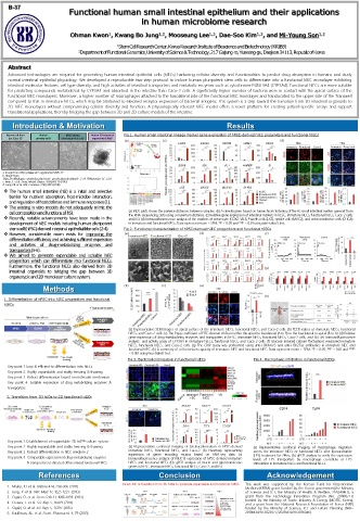
Human cell-lines Human hPSC-deived Human 3D intestinal Fig 1. Human small intestinal lineage marker gene expression of hPSC-derived hIEC progenitors and functional hIECs
(ex. Caco-2) a primary cells b enterocyte-like cells c organoinds (hIOs) d
(a) (c) (d)
a. Image from https://www.atcc.org/products/htb-37 (b)
b. Image from
https://cellbiologics.com/index.php?route=product/product&path=2_50_110&product_id=2373
c. Iwao, T. et al. Drug. Metab. Dispos. 43(4):603-10 (2015)
d. Jung, KB. et al. Nat. Commun. 2;9(1):3039 (2018)
The human small intestine (hSI) is a initial and selective
barrier for nutrient absorption, host-microbe interaction,
andregulationofhostdefenseandimmuneresponses(1).
The existing in vitro models do not adequately mimic the
cellcompositionandfunctionsofhSI. (a) MDS plots shows the pairwise distances between samples. (b) A dendrogram based on hierarchical clustering of the 85 small intestinal marker geneset from
Recently, notable advancements have been made in the the RNA-sequencing data using a maximum distance. (c) Relative gene expression of intestinal markers in hESC, immature hIECs, functional hIECs, Caco-2 cells,
and hSI. (d) Immunofluorescence analysis of the markers of enterocyte (CDX2, VIL1), Paneth cells (LYZ), goblet cells (MUC2), and enteroendocrine cells (CHGA)
development of hIEC models including human pluripotent in immature and functional hIECs. Data represent mean ± SEM. *P < 0.05 and **P < 0.01 using two-tailed t test.
stemcell(hPSC)-derivedintestinalepithelial-likecells(2-4). Fig 2. Functional characterization of hPSC-derived hIEC progenitors and functional hIECs
However, considerable room exists for improving the (a) (d) (f)
differentiationefficiencyandachievingsufficientexpression
and activities of drug-metabolizing enzymes and
transporters(5-6).
We aimed to generate expandable and scalable hIEC
progenitors which can differentiate into functional hIECs.
Furthermore, the functional hIECs also derived from 3D (g)
intestinal organoids to bridging the gap between 3D
organotypicand2Dmonolayerculturesystem.
Methods (b) (c) (e)
(h)
1. Differentiation of hPSC into hIEC progenitors and functional
hIECs
(a) Representative SEM images of apical surface of the immature hIECs, functional hIECs, and Caco-2 cells. (b) TEER values of immature hIECs, functional
hIECs, and Caco-2 cells. (c) The Papp coefficient of FITC-dextran 4 kDa in either the apical to basolateral (A to B) or the basolateral to apical (B to A). (d) Relative
gene expression of drug-metabolizing enzymes and transporters in hESC, immature hIECs, functional hIECs, Caco-2 cells, and hSI. (e) Immunofluorescence
analysis and activity assay of CYP3A4 in immature hIECs, functional hIECs, and Caco-2 cells. (f) Glucose-induced calcium fluctuations measured immature
hIECs, functional hIECs, and Caco-2 cells. (g) The ChIP assay was performed using anti-H3K4me3 and anti-H3K27ac antibodies in immature hIEC and
functional hIEC. (h) A summary of cell retention capacity of immature hIEC and functional hIEC. Data represent mean ± SEM. *P < 0.05, **P < 0.01 and ***P
< 0.001 using two-tailed t test.
Fig 3. Bacterial colonization in functional hIECs Fig 4. Macrophage infiltration in functional hIECs
Key point 1. Easy & efficient to differentiation into hIECs
Key point 2. Highly expandable and stably freezing & thawing
Key point 3. Robust differentiation based on molecular mechanism
Key point 4. Suitable expression of drug-metabolizing enzymes &
transporters
2. Transition from 3D hIOs to 2D functional hIECs
Key point 1. Establishment of expandable 3D InS exp culture system
Key point 2. Highly expandable and stably freezing & thawing (a) Representative conforcal imaging of bactriacolonization in hPSC-derived (a) Representative conforcal imaging of macrophage migration
Key point 3. Robust differentiation in hIEC medium 2 immature hIECs, functional hIECs, and Caco-2. (b) Heatmap representing across the immature hIECs or functional hIECs after liposaccharide
expression of genes encoding mucins based on RNA-seq data. (c)
Key point 4. Comparable expression of drug-metabolizing enzymes Immunofluorescence analysis of MUC13 expression of hPSC-derived immature (LPS) treatment for 24hrs. (b) qPCR analysis to verify the expression
levels of LPS transporters by macrophage co-culture or LPS
& transporters to directed differentiated functional hIECs hIECs and functional hIECs (D) qPCR analysis of mucin and glycotransferase stimulation in immature hIECs and functional hIECs.
genes in hESC, immature hIECs, functional hIECs, Caco-2, and hSI.
References Conclusion Acknowledgement
Direct diif. & transition from 3D hIOs to generate expandable and functional hIECs This work was supported by the Korean Fund for Regenerative
1. Maloy, KJ. et al. Nature 474, 298-306 (2011) Medicine(KFRM) grant funded by the Korea government(the Ministry
2. Jung, P. et al. Nat. Med. 17, 1225-1227 (2013) of Science and ICT, the Ministry of Health & Welfare, 21A0404L1), a
3. Ogaki, O. et al. Stem Cells 31, 1086-1096 (2013) grant from the Technology Innovation Program (No. 20008777)
4. Ozawa, T. et al. Sci. Rep. 5, 16479 (2015) funded by the Ministry of Trade, Industry & Energy (MOTIE, Korea),
and a grant from the National Research Foundation of Korea (NRF)
5. Ogaki, O. et al. Sci. Rep. 5, 17297 (2015) funded by the Ministry of Science, ICT and Future Planning (NRF-
6. Kauffman, AL. et al. Front. Pharmacol. 4, 79 (2013) 2018M3A9H3023077/2021M3A9H3016046).

