


Page 3 - ebook
P. 3
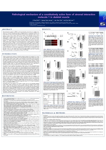
Pathological mechanism of a constitutively active form of stromal interaction
molecule 1 in skeletal muscle
Ji Hee Park 1,2,† , Seung Yeon Jeong 1,2,† , Jun Hee Choi 1,2 and Eun Hui Lee 1,2,*
1 Department of Physiology, College of Medicine, The Catholic University of Korea, Seoul 06591, Korea
2 Department of Biomedicine & Health Sciences, Graduate School, The Catholic University of Korea, Seoul 06591, Korea
† Equal contribution
* correspondence (ehui@catholic.ac.kr)
A B S T RACT R E S ULTS
Stromal interaction molecule 1 (STIM1) is the main protein that, along with Orai1, mediates store-
Control
wt STIM1
operated Ca 2+ entry (SOCE) in skeletal muscle. Abnormal SOCE due to mutations in STIM1 is one 1.00 ± 0.29 1.07 ± 0.35 STIM1-R304Q
8.00 ± 0.27 * ,#
of the causes of human skeletal muscle diseases. STIM1-R304Q (a constitutively active form of Basal SOCE (47) (51) (50)
STIM1) has been found in human patients with skeletal muscle phenotypes such as muscle ▲ Supplementary Table S1. Extracellular Ca 2+ entry
weakness, myalgia, muscle stiffness, and contracture. However, the pathological mechanism(s) of without the depletion of the SR in wtSTIM1 or STIM1-
STIM1-R304Q in skeletal muscle have not been well studied. To examine the pathological R304Q-expressing myotubes. Extracellular Ca 2+ entry
without the depletion of the SR (i.e., basal SOCE) in
mechanism(s) of STIM1- R304Q in skeletal muscle, STIM1-R304Q was expressed in mouse wtSTIM1- or STIM1-R304Q-expressing myotubes was
primary skeletal myotubes, and the properties of the skeletal myotubes were examined using single- measured. The myotubes were incubated with the imaging
solution without Ca 2+ (0 mM) for 15 min, and extracellular
myotube Ca 2+ imaging, transmission electron microscopy (TEM), and biochemical approaches. Ca 2+ (2 mM) was then applied to the myotubes to induce
STIM1-R304Q did not interfere with the terminal differentiation of skeletal myoblasts to myotubes basal SOCE. The experimental mean values were
normalized to the mean values of the control (for the area
and retained the ability of STIM1 to attenuate dihydropyridine receptor (DHPR) activity. STIM1- under the peaks of basal SOCE) and the values are presented
R304Q induced hyper-SOCE (that exceeded the SOCE by wild-type STIM1) by affecting both the as the mean ± SE for the number of myotubes shown in
parentheses. *Significant difference compared with control
amplitude and the onset rate of SOCE. Unlike that by wild-type STIM1, hyper-SOCE by STIM1- (p < 0.05). # Significant difference compared with wtSTIM1
R304Q contributed to a disturbance in Ca 2+ distribution between the cytosol and the sarcoplasmic (p < 0.05).
reticulum (SR) (high Ca 2+ in the cytosol and low Ca 2+ in the SR). Moreover, the hyper-SOCE and
the high cytosolic Ca 2+ level induced by STIM1-R304Q involve changes in mitochondrial shape.
Control
wtSTIM1
Therefore, a series of these cellular defects induced by STIM1-R304Q could induce deleterious ▲ Figure 1. Schematic of the primary structure of STIM1, the expression of RyR1 1.00 ± 0.00 1.01 ± 0.02 STIM1-R304Q
1.01 ± 0.02
skeletal muscle phenotypes in human patients carrying STIM1-R304Q. wtSTIM1 or STIM1-R304Q in mouse primary skeletal myotubes, and myotube DHPR 1.00 ± 0.00 1.05 ± 0.06 1.03 ± 0.04
width. (A) the location of R304 is indicated. Numbers indicate the amino acid SERCA1a 1.00 ± 0.00 1.01 ± 0.02 1.01 ± 0.03
sequence. Each domain of STIM1 is present [7]. S, signal peptide; cEF, canonical EF- Orai1 1.00 ± 0.00 1.02 ± 0.04 0.76 ± 0.04 * ,#
hand; hEF, nonfunctional hidden EF-hand; SAM, sterile α-motif; T, transmembrane STIM1 1.00 ± 0.00 1.01 ± 0.05 1.00 ± 0.03
domain; CC, coiled-coil domian; CAD/SOAR, Ca 2+ release-activated Ca 2+ -activating ▲ Supplementary Table S2. Expression levels of
I N T R O DUC TI O N domain/STIM1-Orai1-activating region; PS, proline/serine-rich domian; and L, lysin- proteins that mediate Ca 2+ movements in wtSTIM1 or
rich domain; (B) mouse primary skeletal myotubes that were transfected with cDNA
of empty vector (control), wtSTIM1, or STIM1-R304Q were stained with anti-GFP STIM1-R304Q-expressing myotubes. The lysate of
wtSTIM1- or STIM1-R304Q-expressing myotubes was
(for detecting CFP or CFP-tagged proteins) and Cy3-conjugated secondary antibodies.
Initiation of skeletal muscle contraction is mediated by excitation–contraction (EC) coupling [1–3]. In short, The bar represents 100µm; (C) myotube width was measured. The mean values of subjected to immunoblot assays with one of the antibodies
against five proteins that mediate intracellular Ca 2+ -release
t-tubule membrane depolarization serially activates (1) the dihydropyridine receptor (DHPR) on the t-tubule each normalized to the mean value of the control are summarized as histograms (Table or SOCE in skeletal muscle. α-actin was used as a loading
1).
membrane, (2) the ryanodine receptor 1 (RyR1) on the sarcoplasmic reticulum (SR) membrane (by physical ▲ Figure 2. Ca 2+ entry through the SOCE mechanism and intracellular control. Three independent experiments were conducted per
each protein. *Significant difference compared with the
interactions between active DHPR and RyR1), and (3) Ca 2+ release from the SR to the cytosol through active Ca 2+ release. (A) Ca 2+ of the SR in wtSTIM1- or STIM1-R304Q-expressing control (p < 0.05). # Significant difference compared with
RyR1. Finally, Ca 2+ in the cytosol turns on a series of contractile proteins by binding to troponin C. Therefore, myotubes was depleted by treatment with thapsigargin (2.5 µM) in the absence wtSTIM1 (p < 0.05).
of extracellular Ca 2+ , and extracellular Ca 2+ (2 mM) was then applied to the
the change in intracellular Ca 2+ levels is a messenger event that connects membrane depolarization to muscle myotubes to induce SOCE. The experimental mean values were normalized to
contraction during skeletal muscle contraction. In addition to the initiation of skeletal muscle contraction, the the mean values of the control (for the area under the peaks on the left-hand
side and for the slope in the rising phase of SOCE on the right-hand side, Table
maintenance of high cytosolic Ca 2+ levels during skeletal muscle contractions, such as long periods or 1). KCl (B) or caffeine (C) was applied to the myotubes, and intracellular Ca 2+ Control wtSTIM1 STIM1-R304Q
repetitive contractions, is another important issue to understand skeletal muscle contraction. release from the SR to the cytosol through RyR1 was measured. The JP1 1.00 ± 0.00 0.99 ± 0.04 1.03 ± 0.04
experimental values were normalized to the mean values of the control (Table JP2 1.00 ± 0.00 0.99 ± 0.03 1.03 ± 0.05
Extracellular Ca 2+ entry contributes to the maintenance of high cytosolic Ca 2+ levels during skeletal muscle 1). A representative trace for each group is shown (A-C). *Significant TRPC1 1.00 ± 0.00 1.04 ± 0.06 1.58 ± 0.12 * ,#
contractions, and store-operated Ca 2+ entry (SOCE) is the main extracellular Ca 2+ entry method in skeletal difference compared with the control (p < 0.05). # Significant difference TRPC3 1.00 ± 0.00 1.03 ± 0.06 1.05 ± 0.06
compared with wtSTIM1 (p <0.05).
muscle [2,3]. Orai1 (a Ca 2+ entry channel) on the t-tubule membrane and stromal interaction molecule 1 ▲ Supplementary Table S3. Expression levels of JP1,
(STIM1, a Ca 2+ sensor) on the SR membrane are the main SOCE-mediating proteins in skeletal muscle by the JP2, TRPC1, or TRPC3 in wtSTIM1 or STIM1-R304Q-
expressing myotubes. The lysate of wtSTIM1- or STIM1-
formation of puncta (i.e., oligomeric complexes of STIM1s and Orai1s). Various mutations in STIM1 (at H72, R304Q-expressing myotubes was subjected to immunoblot
N80, G81, D84, S88, L92, L96, Y98, F108, H109, I115, E136X, P165, L251, R304, R426, R429, and I484) assays with one of the antibodies against JP1, JP2, TRPC1,
have been reported [4–8]. Patients with skeletal muscle diseases involving STIM1 mutations have also been or TRPC3. α-actin was used as a loading control. Three
independent experiments were conducted per each protein.
reported [2–8]. For example, congenital myopathies have been associated with E136X; muscular hypotonia *Significant difference compared with the control (p < 0.05).
with R429C; tubular aggregate myopathy with N80T, G81D, L96V, F108I, H109R, I115F, or I484R; skeletal # Significant difference compared with wtSTIM1 (p < 0.05).
muscle atrophy and progressive muscle weaknesses with H72Q, D84G, H109N, H109R, or R304W.
STIM1 R304 is located in a coiled-coil domain of STIM1, and human patients with substitution of the
arginine at residue 304 by tryptophan (STIM1-R304W, a constitutively active form of STIM1) show
Stormorken syndrome, which is a multisystemic disease characterized by skeletal muscle phenotypes,
including tubular aggregate myopathy, muscle spasms, muscle weakness, atrophy, lack of endurance, and
hematological phenotypes [5,9–11]. Studies on STIM1-R304W with cells from patients with Stormorken
syndrome or model mice carrying STIM1-R304W suggest that excessive SOCE is a cause of multisystemic
defects caused by STIM1-R304W [4,5,12,13]. ▲ Figure 3. Cytosolic Ca 2+ levels, the amount of Ca 2+ releasable from the SR, and
expression levels of Ca 2+ movement-mediating proteins. (A) cytosolic Ca 2+ levels at
Interestingly, patients with substitution of the arginine at residue 304 by the less hydrophobic glutamine rest were measured in wtSTIM1- or STIM1-R304Q-expressing myotubes, and the ▲ Figure 4. Shapes of mitochondria. (A) mitochondria of wtSTIM1- or
(STIM1-R304Q, another constitutively active form of STIM1) rather than tryptophan (STIM1-R304W) mean values are summarized as histograms (Table 1); (B) amount of Ca 2+ releasable STIM1-R304Q-expessing myotubes were observed using TEM. Mitochondria
from the SR to the cytosol was measured in the myotubes by treatment with
manifest a milder and lesser deleterious clinical phenotype of Stormorken syndrome, such as muscle thapsigargin (2.5 µM) in the absence of extracellular Ca 2+ . The mean values of each with concentrically laminated bodies (enlarged images) or long mitochondria
(indicated by arrows) were found in STIM1-R304Q-expressing myotubes. The
weakness, myalgia, muscle stiffness, and contractures [4,9,10]. However, despite these clear symptoms in the normalized to the mean value of the control are summarized as histograms (Table 1); bar represents 2 µm; (B) mitochondrial length was measured. The mean values
skeletal muscle of patients with STIM1-R304Q [4,5,9,10], studies on pathological mechanisms of STIM1- (C) the lysate of the myotubes was subjected to immunoblot assays with antibodies of each normalized to the mean value of the control are summarized as
against five proteins. α-actin was used as a loading control. The expression level of
R304Q have been conducted using only “nonmuscle cells”, such as fibroblasts or a heterologous expression each protein normalized to the value of each control is presented as histograms histograms (Table 1). *Significant difference compared with the control (p
< 0.05). # Significant difference compared with wtSTIM1 (p < 0.05).
system (HEK293 cells) [5,10]. Therefore, in this study, we aimed to reveal the pathological role(s) of STIM1- (Supplementary Table S2). *Significant difference compared with control (p <0.05).
# Significant difference compared with wtSTIM1 (p < 0.05).
R304Q in skeletal muscle at the cellular level using mouse primary skeletal myotubes (instead of a
heterologous expression system involving variations in the expression), single-myotube Ca 2+ imaging
experiments, and biochemical approaches.
Control wt STIM1 STIM1-R304Q
1.00 ± 0.04 1.02 ± 0.05 1.03 ± 0.07
Width of myotubes
(50) (50) (50)
R E FE RE N CE S Peak area 1.00 ± 0.12 1.40 ± 0.11 * 1.88 ± 0.14 * ,#
(40)
(40)
(40)
SOCE
Slope 1.00 ± 0.05 0.99 ± 0.05 1.11 ± 0.06 * ,#
(30) (30) (30)
1. Lee, E.H. Ca 2+ channels and skeletal muscle diseases. Prog. Biophys. Mol. Biol. 2010, 103, 35–43.Lee, E. H. Ca 2+ channels and skeletal muscle diseases. KCl response 1.00 ± 0.05 0.78 ± 0.07 * 0.75 ± 0.06 * ▲ Supplementary Figure S2. Expression levels of JP1,
(72)
(70)
(70)
Prog. Biophys. Mol. Biol. 103, 35-43 (2010). JP2, TRPC1, or TRPC3 in wtSTIM1 or STIM1-R304Q-
2. Cho, C.H.; Woo, J.S.; Perez, C.F.; Lee, E.H. A focus on extracellularCa 2+ entry into skeletal muscle. Exp. Mol. Med. 49, e378 (2017). Caffeine response 1.00 ± 0.05 1.03 ± 0.05 1.04 ± 0.04 expressing myotubes. (A) Lysate of wtSTIM1- or STIM1-
(70)
(70)
(70)
3. Cho, C.H.; Lee, K.J.; Lee, E.H. With the greatest care, stromal interaction molecule (STIM) proteins verify what skeletal muscle is doing. BMB Rep. 51, 78.16 ± 7.07 82.73 ± 7.14 106.76 ± 7.94 * ,# R304Q-expressing myotubes was subjected to immunoblot
378–387 (2018). Resting [Ca 2+ ] cytosol, nM (50) (50) (50) assays with one of the antibodies against JP1, JP2, TRPC1,
Supplementary Figure S1. Extracellular Ca 2+ entry without the
4. Morin, G.; Biancalana, V.; Echaniz-Laguna, A.; Noury, J.B.; Lornage, X.; Moggio, M.; Ripolone, M.; Violano, R.; Marcorelles, P. Marechal, D.; et al. 1.00 ± 0.11 1.02 ± 0.08 0.77 ± 0.09 * ,# ▲ depletion of the SR in wtSTIM1 or STIM1- R304Q-expressing myotubes. or TRPC3. α-actin was used as a loading control. (B) The
Tubular aggregate myopathy and Stormorken syndrome: Mutation spectrum and genotype/phenotypecorrelation. Hum. Mutat. 41, 17–37 (2020) Amount of Ca 2+ releasable from the SR (40) (40) (40) expression level of each protein normalized to the value of
5. Fahrner, M.; Stadlbauer, M.; Muik, M.; Rathner, P.; Stathopulos, P.; Ikura, M.; Muller, N.; Romanin, C. A dual mechanism promotes switching of the Length of mitochondria 1.00 ± 0.05 0.93 ± 0.07 1.51 ± 0.06 * ,# (A) Extracellular Ca 2+ entry without the depletion of the SR (i.e., basal SOCE) each control is presented as histograms (Supplementary
in wtSTIM1- or STIM1-R304Q-expressing myotubes was measured. The
Stormorken STIM1 R304W mutant into the activated state. Nat. Commun. 9, 825 (2018). (62) (61) (69) Table S3). Three independent experiments were conducted
6. Bohm, J.; Chevessier, F.; Maues De Paula, A.; Koch, C.; Attarian, S.; Feger, C.; Hantai, D.; Laforet, P.; Ghorab, K.; Vallat, J.M.; et al. Constitutive myotubes were incubated with the imaging solution without Ca 2+ (0 mM) for per each protein. *Significant difference compared with
activation of the calcium sensor STIM1 causes tubular-aggregate myopathy. Am. J. Hum. Genet. 92, 271–278 (2013). ▲Table 1. Properties of wtSTIM1 or R304Q-expressing mouse primary skeletal 15 min, and extracellular Ca 2+ (2 mM) was then applied to the myotubes to
7. Choi, J.H.; Huang, M.; Hyun, C.; Oh, M.R.; Lee, K.J.; Cho, C.H.; Lee, E.H. A muscular hypotonia-associated STIM1 mutant at R429 induces myotubes. The values, except for those of the cytosolic Ca 2+ levels at rest, were induce basal SOCE. A representative trace for each group is shown. (B) The control (p < 0.05). # Significant difference compared with
wtSTIM1 (p < 0.05).
abnormalities in intracellularCa 2+ movement and extracellular Ca 2+ entry in skeletal muscle. Sci. Rep. 9, 19140 (2019). normalized to the mean value of those from the control. The values are presented as experimental mean values were normalized to the mean values of the control
8. Lee, K.J.; Woo, J.S.; Hwang, J.H.; Hyun, C.; Cho, C.H.; Kim, D.H.; Lee, E.H. STIM1 negatively regulates Ca 2+ release from the sarcoplasmic reticulum the mean ± SE for the number of myotubes shown in parentheses. *Significant (for the area under the peaks of basal SOCE) and the values are presented as
in skeletal myotubes. Biochem. J. 453, 187–200 (2013). difference compared with control (p < 0.05). # Signiticant difference compared with the mean ± SE for the number of myotubes shown in parentheses of
9. Borsani, O.; Piga, D.; Costa, S.; Govoni, A.; Magri, F.; Artoni, A.; Cinnante, C.M.; Fagiolari, G.; Ciscato, P.; Moggio, M.; et al. Stormorken Syndrome wtSTIM1 (p < 0.05). Supplementary Table S1. *Significant difference compared with control (p
Caused by a p.R304W STIM1 Mutation:The First Italian Patient and a Review of the Literature. Front. Neurol. 9, 859 (2018). < 0.05). # Significant difference compared with wtSTIM1 (p < 0.05).
10. Harris, E.; Burki, U.; Marini-Bettolo, C.; Neri, M.; Scotton, C.; Hudson, J.; Bertoli, M.; Evangelista, T.; Vroling, B.; Polvikoski, T.; et al. Complex
phenotypes associated with STIM1 mutations in both coiled coil and EF-hand domains. Neuromuscul. Disord. 27, 861–872(2017).
11. Silva-Rojas, R.; Laporte, J.; Bohm, J. STIM1/ORAI1 Loss-of-Function and Gain-of-Function Mutations Inversely Impact on SOCE and
Calcium Homeostasis and Cause Multi-Systemic Mirror Diseases. Front. Physiol. 11, 604941 (2020).
12. Misceo, D.; Holmgren, A.; Louch, W.E.; Holme, P.A.; Mizobuchi, M.; Morales, R.J.; De Paula, A.M.; Stray-Pedersen, A.; Lyle, R.; Dalhus, B.; M AT E RIAL S & M E T H O D S
et al.A dominant STIM1 mutation causes Stormorken syndrome. Hum. Mutat. 35, 556–564 (2014).
13. Gamage, T.H.; Gunnes, G.; Lee, R.H.; Louch, W.E.; Holmgren, A.; Bruton, J.D.; Lengle, E.; Kolstad, T.R.S.; Revold, T.; Amundsen, S.S.; et al.
STIM1 R304W causes muscle degeneration and impaired platelet activation in mice. Cell Calcium.76, 87–100 (2018).
14. Rando, T.A.; Blau, H.M. Methods for myoblast transplantation.Methods Cell Biol. 52, 261–272 (1997). Ethical Approval
The methods were carried out in accordance with the guidelines and regulations of the College of Medicine at the Catholic University of Korea. All surgical interventions, including pre- and postsurgical animal care and the site where the
15. Lee, K.J.; Park, C.S.; Woo, J.S.; Kim, D.H.; Ma, J.; Lee, E.H. Mitsugumin 53 attenuates the activity of sarcoplasmic reticulum Ca 2+ -ATPase 1a animal work was performed, were carried out in accordance with the Laboratory Animals Welfare Act, the Guide for Care and Use of Laboratory Animals, and the Guidelines and Policies for Rodent Survival Surgery approved by the
(SERCA1a) in skeletal muscle. Biochem.Biophys.Res. Commun. 428, 383–388 (2012).
16. Woo, J.S.; Cho, C.H.; Lee, K.J.; Kim, D.H.; Ma, J.; Lee, E.H. Hypertrophy in skeletal myotubes induced by junctophilin-2 mutant, Y141H, Institutional Animal Care and Use Committee of the College of Medicine at the Catholic University of Korea. All experimental protocols were approved by the Committee of the College of Medicine at the Catholic University of Korea
involves an increase in store-operated Ca 2+ entry via Orai1. J. Biol.Chem. 287, 14336–14348 (2012). (2017-0117-01).
17. Lee, K.J.; Hyun, C.; Woo, J.S.; Park, C.S.; Kim, D.H.; Lee, E.H. Stromal interaction molecule 1 (STIM1) regulates sarcoplasmic/endoplasmic cDNA Construction, Cell Culture, and STIM1-R304Q Expression
reticulum Ca 2+ -ATPase 1a (SERCA1a) in skeletal muscle. Pflugers Arch. 466, 987–1001(2014).
18. Ahn, M.K.; Lee, K.J.; Cai, C.; Huang, M.; Cho, C.H.; Ma, J.; Lee, E.H. Mitsugumin 53 regulates extracellular Ca 2+ entry and intracellular Ca 2+ The mutation of the R at residue 304 of STIM1 to Q (STIM1-R304Q) was carried out using human STIM1 cDNA as a template (GenBank accession number: NM_003156.3), a site-directed mutagenesis kit (Agilent Technologies, Santa Clara,
CA, USA), and a pair of complementary synthetic oligonucleotide primers containing the desired mutation (forward primer, 5’-CGGCTGAAGGAGCTGCAGGAGGGTACTGAGAATG-3’; reverse primer, 5’-
release via Orai1 and RyR1 in skeletal muscle. Sci. Rep. 6, 36909 (2016).
19. Oh, M.R.; Lee, K.J.; Huang, M.; Kim, J.O.; Kim, D.H.; Cho, C.H.; Lee, E.H. STIM2 regulates both intracellular Ca 2+ distribution and Ca 2+ CATTCTCAGTACCCTCCTGCAGCTCCTTCAGCCG-3’) [7,8]. Mouse primary skeletal myoblasts were derived from mouse skeletal muscle using a single-cell cloning method, expanded, and differentiated into myotubes, as previously
movement in skeletal myotubes. Sci. Rep. 7, 17936 (2017). described [7,8,14,15]. After three days of culture under differentiation conditions, premature myotubes were transfected with an empty vector or cDNA encoding wtSTIM1 or STIM1-R304Q for 3 h. Mature myotubes were imaged, observed,
20. Huang, M.; Lee, K.J.; Kim, K.J.; Ahn, M.K.; Cho, C.H.; Kim, D.H.; Lee, E.H. The maintenance ability and Ca 2+ availability of skeletal muscle or disrupted at 36 h posttransfection for further experiments. All reagents for the cell cultures were obtained from Invitrogen (Thermo Fisher Scientific, Waltham, MA, USA).
are enhanced by sildenafil. Exp. Mol. Med. 48, e278 (2016). Immunocytochemistry and Immunoblot Assays
21. Silva-Rojas, R.; Treves, S.; Jacobs, H.; Kessler, P.; Messaddeq, N.; Laporte, J.; Bohm, J. STIM1 over-activation generates a multisystemic For immunocytochemistry assays, myotubes were fixed in cold methanol (−20 °C) for 30 min and permeabilized with 0.05% Tween 20 in PBS for 1 min, as previously described [7,8,16,17]. For immunoblot assays, solubilized lysate of
phenotypeaffecting the skeletal muscle, spleen, eye, skin, bones and immune system in mice. Hum. Mol. Genet. 28, 1579–1593(2019).
22. Zhou, Y.; Wang, X.; Wang, X.; Loktionova, N.A.; Cai, X.; Nwokonko, R.M.; Vrana, E.; Wang, Y.; Rothberg, B.S.; Gill, D.L. STIM1 dimers undergo myotubes (15 µg of total protein) was subjected to SDS–PAGE (8 or 10% gel), as previously described [7,8,16–20]. Anti-RyR1 and anti-SERCA1a antibodies were obtained from Affinity BioReagents. Anti-DHPR, anti-Orai1, anti-STIM1,
unimolecular coupling to activate Orai1 channels. Nat. Commun. 6, 8395 (2015). and anti-α-actin antibodies were obtained from Abcam (Cambridge, MA, USA).
23. Nesin, V.; Wiley, G.; Kousi, M.; Ong, E.C.; Lehmann, T.; Nicholl, D.J.; Suri, M.; Shahrizaila, N.; Katsanis, N.; Gaffney, P.M.; et al. Activating Single-Myotube Ca 2+ Imaging
mutations in STIM1 and ORAI1 cause overlapping syndromes of tubular myopathy and congenital miosis. Proc. Natl. Acad. Sci. USA 111, Single-myotube Ca 2+ imaging was performed using an inverted-stage microscope (Nikon Eclipse TS100, Nikon Instruments, Inc., Melville, NY, USA) and a high-speed monochromator with a 75 W xenon lamp (FSM150Xe, Bentham
4197–4202 (2014).
24. Park, C.Y.; Shcheglovitov, A.; Dolmetsch, R. The CRAC channel activator STIM1 binds and inhibits L-type voltage-gated calcium channels. Instruments, Reading, Berkshire, UK), as previously described [7,8,16–18,20]. The data were analyzed using image acquisition and analysis software (High-Speed InCyt Im1 and Im2, v5.29, Intracellular Imaging Inc., Cincinnati, OH, USA).
Slopes at the rising phase of SOCE were examined by a linear equation, as previously described [7]. Reagents for single-myotube Ca 2+ imaging were obtained from Sigma-Aldrich (St. Louis, MO, USA).
Science, 330, 101–105 (2010).
25. des Georges, A.; Clarke, O.B.; Zalk, R.; Yuan, Q.; Condon, K.J.; Grassucci, R.A.; Hendrickson, W.A.; Marks, A.R.; Frank, J. Structural Basis Transmission Electron Microscopy (TEM) Observation, Myotube Width Measurement, and Mitochondrial Length Measurement
for Gating and Activation of RyR1. Cell, 167, 145–157.e117(2016). Myotubes were fixed, embedded in epoxy resin (Epon 812), sectioned (70–80 nm) using an ultramicrotome (Ultracut UCT ultramicrotome, Leica, Buffalo Grove, IL, USA), and examined under TEM (JEM1010, JEOL Ltd., Peabody, MA,
26. Choi, J.H.; Jeong, S.Y.; Oh, M.R.; Allen, P.D.; Lee, E.H. TRPCs: Influential Mediators in Skeletal Muscle. Cells, 9, 850 (2020). USA), as previously described [7,16]. Myotube width measurements or mitochondrial length measurements were performed using ImageJ software, as previously described [7,8,16,17,20].
27. Neustein, H.B.; Lurie, P.R.; Dahms, B.; Takahashi, M. An X-linked recessive cardiomyopathy with abnormal mitochondria. Pediatrics, 64, 24–
29 (1979). Statistical Analysis
28. Hood, D.A.; Memme, J.M.; Oliveira, A.N.; Triolo, M. Maintenance of Skeletal Muscle Mitochondria in Health, Exercise, and Aging. Annu. Rev. Physiol. The results are presented as the mean ± SE for the number of myotubes shown in parentheses in the figure legends or tables. Group differences were analyzed using an unpaired t-test (GraphPad InStat, v2.04, GraphPad Software, San Diego,
81, 19–41 (2019). CA, USA). The differences were considered to be significant at p < 0.05. The graphs were prepared using Origin 2019b (OriginLab, Northampton, MA, USA).

