


Page 65 - ebook
P. 65
Nrf2 activation by 2-methoxycinnamaldehyde attenuates macrophage-
                             proinflammatory responses via enhancing autophagy flux


                                    Minwook Shin 1,# , Bo-Sung Kim 2,3,# , Jin-Yeong Kim 2,3 , Ki-Tae Ha 2,3 , Sung-Jin Bae 3,*
                                 1 RNA Therapeutics Institute, University of Massachusetts Medical School, Worcester, MA, USA
                        2 Department of Korean Medical Science, School of Korean Medicine, Pusan National University, Yangsan, Gyeongnam 50612, Korea
                            3 Korean Medical Research Center for Healthy Aging, Pusan National University, Yangsan, Gyeongnam 50612, Korea

                ABSTRACT                                          RESULTS

       Inflammation is the body's response to infection, tissue  Fig. 1. Anti-inflammatory effects of 2-MCA in LPS-  Fig. 2. Effect of 2-MCA on activation of MAPK,
       damage,  and  cellular  stress.  A  well-controlled  stimulated macrophages.  NF-kB, and AP-1 pathways in RAW264.7 and
       inflammatory response has a beneficial effect for                 BMMs.
       recovery and maintainance of tissue homeostasis. 2-
       methoxycinnamaldehyde  (2-MCA)  is  a  natural
       compound found in cinnamon, the most common
       spice. The anti-inflammatory response of 2-MCA has
       been  already  known;  however,  the  mechanism
       underlying anti-inflammatory action was not fully
       elucidated. It was confirmed that the LPS-stimulated
       production of TNF-α and NO was reduced by 2-MCA
       treatment in murine bone marrow macrophages
       (BMM) and macrophagic RAW264.7 cells. To
       investigate the mechanism by which 2-MCA has anti-
       inflammatory effects, MAPK, NF-κB, and AP-1
       signaling pathways activated by LPS were examined.
       2-MCA treatment did not affect the MAPK, NF-κB,
       and AP-1 signaling pathways. However, 2-MCA
       increased the expression and nuclear localization of
       Nrf2 in macrophages. Previous studies have suggested
       that Nrf2 is associated with regulation of autophagy.
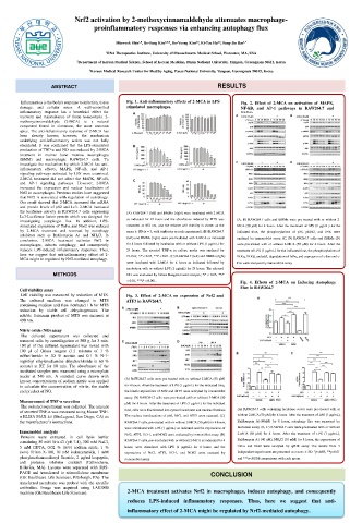
       Our result showed that 2-MCA increased the mRNA
       and protein levels of p62 and Lc3. 2-MCA increased
       the luciferase activity in RAW264.7 cells expressing  (A) RAW264.7 (left) and BMMs (right) were incubated with 2-MCA
       Lc3-Luciferase fusion protein which was designed for
       investigating autophagic flux. In addition, LPS-  as indicated for 24 hours and the absorbance induced by MTS was  (A, B) RAW264.7 cells and BMMs were pre-treated with or without 2-
       stimulated expression of Tnf-α and Nos2 was reduced  measured at 492 nm, and the relative cell viability is shown as the  MCA (50 μM) for 4 hours. After the treatment of LPS (1 μg/mL) for the
       by 2-MCA treatment and reversed by autophagy  mean ± SD (n = 3, with triplicates in each experiment). (B) RAW264.7  indicated time, the phosphorylations of p38, p44/42, and JNK were
       inhibitors such as Bafilomycin A1 and NH 4 Cl. In  (left) and BMMs (right) were pre-incubated with 2-MCA as indicated
       conclusion, 2-MCA treatment activates Nrf2 in                    analyzed by immunoblot assay. (C, D) RAW264.7 cells and BMMs (D)
       macrophages, induces autophagy, and consequently  for 4 hours followed by incubation with or without LPS (1 μg/mL) for  were pre-treated with or without 2-MCA (50 μM) for 4 hours. After the
       reduces LPS-induced inflammatory responses. Thus,  24 hours. The secreted TNF-α in culture media was analyzed by  treatment of LPS (1 μg/mL) for the indicated time, the phosphorylations of
       here we suggest that anti-inflammatory effect of 2-  ELISA; *P < 0.05, **P < 0.01. (C) RAW264.7 (left) and BMMs (right)
       MCA might be regulated by Nrf2-mediated autophagy.               IKKα, IKKβ, and p65, degradation of IκBα, and expression of c-Jun and c-
                                       were incubated with 2-MCA for 4 hours as indicated followed by  Fos were analyzed by immunoblot assay.
                                       incubation with or without LPS (1 μg/mL) for 24 hours. The released
                 METHODS               NO was evaluated by Griess Reagent-based analysis; *P < 0.05, **P
                                       < 0.01, ***P < 0.001.
                                                                         Fig. 4. Effects of 2-MCA on Inducing Autophagy
                                                                         Flux in RAW264.7
       Cell viability assay
       Cell viability was measured by reduction of MTS.  Fig. 3. Effect of 2-MCA on expression of Nrf2 and
       The cultured medium was changed to MTS  ATF3 in RAW264.7.
       containing medium and then incubated 1 h for MTS
       reduction by viable cell dehydrogenases. The
       soluble formazan product of MTS was measure at
       490 nm.
       Nitric oxide (NO) assay
       The cultured supernatant was collected and
       removed cells by centrifugation at 500 g for 3 min.
       100 μl of the cultured supernatant was mixed with
       100 μl of Griess reagent (1:1 mixture of 1 %
       sulfanilamide in 30 % acetate and 0.1 % N-1-
       naphthyl ethylenediamine dihydrochloride in 60 %
       acetate) at RT for 10 min. The absorbance of the
       incubated samples was measured using a microplate
       reader at 540 nm. A standard curve drawn with  (A) RAW264.7 cells were pre-treated with or without 2-MCA (50 μM)
       known concentrations of sodium nitrite was applied
       to calculate the concentration of nitrite, the stable  for 4 hours. After the treatment of LPS (1 μg/mL) for the indicated time,
       end product of NO.             the kinetic expressions of Nrf2 and ATF3 were analyzed by immunoblot
                                      assay. (B) RAW264.7 cells were pre-treated with or without 2-MCA (50
       Measurement of TNF-α secretion  μM) for 4 hours. After the treatment of LPS (1 μg/mL) for the indicated
       The cultured supernatant was collected. The amount
       of secreted TNF-α was measured using Mouse TNF-  time, cells were fractionated into cytosol/membrane and nucleus fractions.  (A) RAW264.7 cells containing luciferase vector were pre-treated with or
       α ELISA MAX kit (BioLegend, San Diego, CA) as  The nucleus translocations of p65, Nrf2, and ATF3 were assessed. (C)  without 2-MCA (50 μM) for 4 hours. After the treatment of LPS (1 μg/mL),
       the manufacturer’s instructions.  RAW264.7 cells, pre-treated with or without 2-MCA (50 μM) for 4 hours,  Bafilomycin A1(40nM) for 8 hours, autophagy flux was measured by
                                      were stimulated with LPS (1 μg/mL) as indicated and the expressions of  luciferase assay. (B, C) RAW264.7 cells were pre-treated with or without
       Immunoblot analysis            Nrf2, ATF3, HO-1, and NOS2 were evaluated by immunoblot assay. (D)  2-MCA (50 μM) for 4 hours. After the treatment of LPS (1 μg/mL),
       Proteins were extracted in cell lysis buffer
       containing 50 mM Tris-Cl (pH 7.4), 300 mM NaCl,  RAW264.7 cells, pre-incubated with or without 2-MCA as indicated for 4  Bafilomycin A1 (40 nM), NH 4 Cl (10 mM) for 8 hours, the expressions of
       5 mM EDTA, 0.02 % (w/v) sodium azide, 1 %  hours, were stimulated with LPS (1 μg/mL) for 4 hours, and the  Tnf-α and Nos2 were analyzed by qPCR assay. The results from 3
       (w/v) Triton X-100, 10 mM iodoacetamide, 1 mM  expressions of Nrf2, ATF3, HO-1, and NOS2 were assessed by  independent experiments are presented as means ± SD. *p<0.05, **p<0.01
       phenylmethanesulfonyl fluoride, 2 μg/ml leupeptin,  immunoblot assay.  and ***p<0.001 comparisons with each group.
       and  protease  inhibitor  cocktail  (Calbiochem,
       Billerica, MA). Lysates were separated with SDS-
       PAGE and transferred to nitrocellulose membrane          CONCLUSION
       (GE Healthcare Life Sciences, Pittsburgh, PA). The
       transferred membrane was probed with the specific
       antibodies. Image was acquired using LAS3000
       machine (GE Healthcare Life Sciences).  2-MCA treatment activates Nrf2 in macrophages, induces autophagy, and consequently
                                       reduces LPS-induced inflammatory responses. Thus, here we suggest that anti-
                                       inflammatory effect of 2-MCA might be regulated by Nrf2-mediated autophagy.

