


Page 61 - ebook
P. 61
Liver-specific deletion of mouse CTCF leads to hepatic steatosis
via augmented PPARγ signaling
1,2
1,2
Woong-Jae Jung , Min-Ji Song , Yeeun Choi 1,2 and Hyoung-Pyo Kim 1
1 Department of Environmental Medical Biology, Institute of Tropical Medicine, Yonsei University College of Medicine, Seoul, Korea
2 These authors contributed equally to this work
Abstract
Background & Aims: The liver is the major organ for metabolizing lipids, and malfunction thereof leads to various diseases. Non-alcoholic fatty liver disease (NAFLD) is rapidly becoming a major health concern worldwide and is characterized
by abnormal retention of excess lipids in the liver. CCCTC-binding factor (CTCF) is a highly conserved zinc finger protein that regulates higher-order chromatin organization and is involved in various gene regulation processes. Here, we sought to
determine the physiological role of CTCF in hepatic lipid metabolism.
Methods: We generated liver-specific CTCF-ablated (cKO) and/or CD36 whole-body knockout mice. Overexpression or knockdown of PPARγ in the liver was achieved using adenovirus. Mice were examined for development of hepatic steatosis
and inflammation. RNA-seq was performed to identify genes affected by CTCF depletion. Genome-wide occupancy of H3K27ac, PPARγ and CTCF were analyzed by chromatin immunoprecipitation sequencing. Genome-wide chromatin
interactions were analyzed by in situ Hi-C.
Results: Liver-specific CTCF-deficient mice (cKO) developed hepatic steatosis and inflammation when fed a standard diet compared to wild type mice (WT). Global analysis of the transcriptome and enhancer landscape revealed that CTCF-
depleted liver exhibited enhanced accumulation of PPARγ in the nucleus, which leads to increased expression of its downstream target genes, including fat storage-related gene CD36, involved in lipid metabolic process. Hepatic steatosis
developed in liver-specific CTCF-deficient mice (cKO) was ameliorated by repression of PPARγ via adenovirus-mediated knockdown, but hardly rescued by additional knockout of CD36.
Result
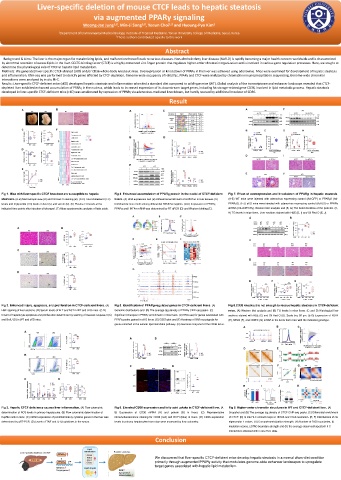
Fig 1. Mice with liver-specific CTCF knockout are susceptible to hepatic Fig 4. Enhanced accumulation of PPARg protein in the nuclei of CTCF-deficient Fig 7. Effect of overexpression and knockdown of PPARg in hepatic steatosis.
steatosis. (A-B) Macroscopic view (A) and Oil Red O staining (B). (C-D) Total cholesterol (TC) livers. (A) RNA expression and (B) Differential enrichment of H3K27ac in liver tissues. (C) (A-E) WT mice were injected with adenovirus expressing control (Ad-GFP) or PPARγ2 (Ad-
levels and triglyceride (TG) levels in liver (C) and serum (D). (E) Plasma TG levels at the Enriched de novo motif among differential H3K27ac regions. (D-E) Expression of PPARγ, PPARγ2). (F-J) cKO mice were injected with adenovirus expressing control (Ad-US) or PPARγ
indicated time points after injection of tyloxapol. (F) Mass spectrometric analysis of fatty acids. PPARα and HNF4α mRNA was determined by RT-qPCR (D) and Western blotting (E). shRNA (Ad-shPPARγ). Western blot analysis and (B, G) The band densities of the proteins. (C,
H) TG levels in mice livers. Liver sections stained with H&E (D, I) and Oil Red O (E, J).
Fig 2. Enhanced injury, apoptosis, and proliferation in CTCF-deficient livers. (A) Fig 5. Identification of PPARg-regulated genes in CTCF-deficient livers. (A) Fig 8.CD36 knockout is not enough to rescue hepatic steatosis in CTCF-deficient
H&E staining of liver sections. (B) Serum levels of ALT and AST in WT and cKO mice. (C-D) Genomic distributions and (B) The average tag density of PPARγ ChIP-seq peaks. (C) mice. (A) Western blot analysis and (B) TG levels in mice livers. (C and D) Histological liver
Extent of hepatocyte apoptosis and proliferation determined by staining of cleaved caspase-3 (C) Significant changes in PPARγ enrichment in mice livers. (D) RNA-seq for genes associated with sections stained with H&E (C) and Oil Red O (D). Scale bar, 50 μm. (E-G) Expression of PDK4
and BrdU (D) in WT and cKO mice. PPARγ peaks gained in cKO livers. (E) GSEA plot and (F) Heatmap of RNA-seq signals for (E), NEU3 (F), and CIDEC (G) mRNA in the livers from mice with the indicated genotype.
genes enriched in the cellular lipid metabolic pathway. (G) Genomic snapshot of the CD36 locus.
Fig 3. Hepatic CTCF deficiency causes liver inflammation. (A) Flow cytometric Fig 6. Elevated CD36 expression and fatty acid uptake in CTCF-deficient liver. (A- Fig 9. Higher-order chromatin structures in WT and CTCF-deficient liver. (A)
determination of ROS levels in primary hepatocytes. (B) Flow cytometric determination of B) Expression of CD36 mRNA (A) and protein (B) in livers. (C) Representative Snapshot and (B) The average tag density of CTCF ChIP-seq peaks. (C) Differential enrichment
Kupffer cells in mice. (C) mRNA expression of proinflammatory cytokine genes in the liver was immunofluorescence staining for CD36 (red) and DAPI (blue) in livers. (D) CD36 expression of CTCF. (D) In situ Hi-C contact maps at 100-kb and 10-kb resolution. (E, F) Distributions of cis
determined by qRT-PCR. (D) Levels of TNF and IL-1β cytokines in the serum. levels in primary hepatocytes from mice were assessed by flow cytometry. eigenvector 1 values. (G) Compartmentalization strength. (H) Number of TAD boundaries, (I)
insulation scores, (J)TAD boundary strength and (K) the average observed/expected Hi-C
interactions obtained with in situ Hi-C data.
Conclusion
We discovered that liver-specific CTCF-deficient mice develop hepatic steatosis in a normal chow-diet condition
primarily through augmented PPARγ activity that modulates genome-wide enhancer landscapes to upregulate
target genes associated with hepatic lipid metabolism.

