


Page 59 - ebook
P. 59
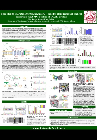
Base editing of Arabidopsis thaliana DGAT1 gene for modification of seed oil
                  biosynthesis and 3D structure of DGAT1 protein
                                Won Nyeong Kimand Hyun Uk Kim*
          Department of Bioindustry and Bioresource Engineering, Sejong University, Seoul, 05006 Republic of Korea

                        Abstract

    In rising concern of climate change and dissipating petroleum,  importance of vegetable oil ( triacylglycerol:TAG) is getting
    highlight.  Due to the structure similarity between petroleum and vegetable oil, many industrial products which traditionally
    processed from petroleum can be replaced with  phyto-based materials. To meet  escalating demands of vegetable oil, appending
    agricultural farm to produce more amount is simple but there are spatial huddles such as region specificity considering growth
    condition of each oil producing plant.  To overcome these issues, increase of vegetable oil producing amount per plant is the  key.
    At this point, diacylglycerol acyltransferase 1 (DGAT1) is crucial enzyme to solve the problem. DGAT1 is endoplasmic reticulu m
    (ER) located transmembrane enzyme and take role in final transfer of acyl chain to diacylglycerol to make TAG.  TAG are
    accumulated between two layers of ER membrane forming monolayer oil body in seeds. In this study, we used base editing
    technology to edit fifty-two target sites in seven functional domains of DGAT1 expecting change of enzyme properties in manner   Fig 4. Changed base of Domain  3 treated
    of increasing TAG produce. By far, total  five type of base changed homo transgenic Arabidopsis were generated. We will   line and its seed fatty acid profile
    discuss the changes in oil composition and content in the seeds of these five DGAT1 variant mutants.
                                                                                          A) Chromatograph result of DNA
                                                                                          Sanger sequencing and substituted
                     Introduction                                                         amino acid occurred by domain 3 base
                                                                                          editing.  B) Alignment  of acyl-CoA
                                                                                          binding motif  (Domain 3). Substituted
                                                                                          amino acid highlighted  with yellow
                                                                                          shade. C) Fatty acid profile of D3-1 lines
                                                                                          using gas chromatography

    Fig 1. Schematic  construction  of DGAT1 domains  and process of Base Editing
    A) Schematic Aradisopsis thatliana DGAT1 structure. Five red colored domains indicating predicted in early report. Remaining
    Fig.  1 Schematic  construction  of DGAT1 domains  and process of Base Editing
    two green colored domains are highly studied its features or investigated by site -directed mutagenesis. Transmembrane
    domains were predicted by TMHMM online tool.  B) Principle of base editing.  Base editor use nick or dead Cas9 which dead or
    weakened on its cleavage activity. When n/dCas9 locate the deaminase domain on target sequence, deaminase domain
    changes the base in window of base editor.
    With this  base editing system, we designed fifty -two targets to induce substitution of amino acids in those domains without   Fig.  5 Changed base of Domain  5 treated line and its seed fatty acid profile
    frame-shift.  As result, five homogeneous Arabidopsis lines were obtained and analyzed by Gas Chromatography(GC) to   A) Chromatograph result of DNA Sanger sequencing and substituted amino acid occurred by domain 3 base editing. B) Alignment  o f
    compare the fatty acid component or amount. The investigation of base edited lines were showed the difference between Col -0   acyl-CoA binding motif  (Domain 5). Substituted  amino acid highlighted with yellow shade. C) Qualitive,  quantitative Fatty acid p rofile of
    wildtype and dgat1 mutant.  But unfortunately, not all targeted domain were substituted to different  amino acid. Furthermore,
    human DGAT1 enzyme, which highly similar in predicted structure with  Ararbidopsis DGAT1,  was reported its 3D structure and   D5-1 and D5-2 lines using gas chromatography
    biochemical features but we will discuss about these issues with our result.
                                                                             Discussion


                                                                                   Fig 6. Predicted  3D structure of DGAT1 based on human
                                                                                   DGAT1 and schematic interaction  of mutant DGAT1 and
                                                                                   acyl-CoA chain.
                                                                                   All 3D models were constructed by Phyer2 web tool that use
                                                                                   homology-based building algorithm. Based on human DGAT1
                                                                                   protein property, Arabidopsis DGAT1 makes dimer form as
                                                                                   normal status and have two main entrance to reaction chamber
                                                                                   which includes Histidine reaction residue. The two main
    Fig 2. Schematic  process of screening  base edited Arabidopsis  and example of base substitution  through generation  entrance is entry tunnel and lateral gate which gives
                                                                                   accessibility to cytosolic region and hydrophobic region of
    To transfer sgRNA including vectors to Arabidopsis, we used floral dipping method  or spraying method to inoculate   membrane, respectively.
    Agrobacterium. After  transformation, harvested T1 seeds were screened by Hygromycin antibiotics. Through these process,   A) 3D model of membrane-imbedded DGAT1 protein.  Lateral
    screened individual plants DGAT1 DNA sequences were send to company for analyze. Hetero individuals which showed mixed   gate had indicated with red dotted circle. B) 3D model of
    pick in sequencing results in T2 or T3 generation were selected to keep breeding. With generation continued, some bases were   domain 1 base edited DGAT1.  Entry tunnel had indicated with
    edited to homogeneous status. Homogeneous lines were analyzed by gas chromatography to investigate the difference between   green dotted circle. Cyan colored residue and line indicate
    Col-0 wildtype and dgat1 mutants.                                              substituted amino acid. Putative N-terminal also extended with
                                                                                   red dotted line. C) 3D model of domain 3 base edited DGAT1.
                                                                                   Red surface indicate substituted amino acid, which has different
                                                                                   binding angle to original one. D) 3D model of domain 5 base
                        Results                                                    edited DGAT1. Amino  acid substitute affects negatively to acyl-
                                                                                   CoA binding indicated with red surface. Amino  acid substitute
                                                                                   affects positively to acyl-CoA binding indicated with green
                                                                                   surface. Entrance of entry tunnel  highligh ted green dotted circle.
                                                                                           Summary
                                                                                   ➢ The  activity  of base edited  DGAT1  seems  weaken
                                                              On-going study       ➢ By 3D  modeling,  edited  regions  are  located.
                                                                                   ➢ Gas Chromatography  shows  tag1-1 similar
                                                         ➢ Comparison  of Gene expression, protein accumulation
                                                          amount                    quantitative  and  qualitative  result  except  20:1  fatty
                                                                                    acid  which  imply  base  edited  AtDGAT1  has
                                                         ➢ Production  of complementary AtDGAT1 transgenic line  different  mode  of action  from  tag1-1 DGAT1
                               Fig 3. Changed base of Domain  1 treated line and its
                               seed fatty acid profile                      References
                               A) Chromatograph result of DNA Sanger sequencing
                               and substituted amino acid occurred by domain 1 base   ➢ Xu, J., Francis, T., Mietkiewska, E.,  Giblin, E.M.,  Barton, D.L.,  Zhang, Y.,  Zhang, M. and Taylor, D.C. (2008), Cloning and
                               editing.  B) Alignment  of acyl-CoA binding motif  (Domain   characterization of an acyl‐CoA‐dependent diacylglycerol acyltransferase 1 (DGAT1) gene from Tropaeolum majus, and a study of
                               1) from various plant species including Homo sapiens.   the functional motifs of the DGAT protein using site‐directed mutagenesis to modify enzyme activity and oil content.  Plant
                               Substitu ted am ino acid high lighted wi th yellow  shade. C)   Biotechnology Journal, 6: 799-818. doi:10.1111/j.1467-7652.2008.00358.x
                               Fatty acid profile of D1-1 and D1-2 lines using gas   ➢ Wang, L., Qian, H., Nian, Y. et al. Structure and mechanism of human diacylglycerol O-acyltransferase 1. Nature 581, 329–332
                               chromatography             (2020). https://doi.org/10.1038/s41586-020-2280-2.
                                     Sejong University, Seoul Korea

