


Page 69 - ebook
P. 69
Andrographolide, a Novel Pyruvate Dehydrogenase Kinase Inhibitor,
                                 Induces Apoptosis of Human Lung Cancer Cells
                                  Yoonju Do 1,2 , EunSun Yang 2 , Jung-Suk Jin 2 , Kibong Kim 3 , Ki-Tae Ha 1,2,*
                       1 Department of Korean Medical Science, School of Korean Medicine, Pusan National University, Yangsan 50612, Korea;
                       2 Korean Medical Research Center for Healthy Aging, Pusan National University, Yangsan 50612, Korea;
                       3 Department of Pediatrics, Pusan National University Korean Hospital, Yangsan 50612, Korea;

               ABSTRACT                                           RESULTS
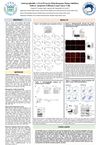
       Most of cancer cells produce their energy                        Figure 2. Andrographolide reduced the lactate
       through a high level of glycolysis followed by  Figure 1. Cell viability assay in selected cell lines.
       lactic acid fermentation even in the presence                    production but increased mitochondrial ROS and
                                                                        mitochondrial damage.
       of  abundant  oxygen.  The  pyruvate
       dehydrogenase kinase (PDK) 1 is commonly
       overexpressed in tumors and recognized as a
       therapeutic target in cancer. Here, we report
       Andrographolide (AG), a diterpenoid lactone
       isolated from Andrographis paniculate, as a
       novel PDK inhibitor through screening from
       natural product library. AG has been reported
       to  anti-inflammatory,  anti-viral,  immune-
       stimulant properties and induced apoptosis in
       cancer  cells.  However,  the  precise
       mechanism of AG induces apoptosis has not
       yet been clearly elucidated. As a result of
       checking the cell viability by MTT assay with
       various cells with drugs, only AG showed
       cytotoxicity correlated with PDK1 expression
       among the 17 types of PDK1 inhibitors
       searched. AG decreases the PDK1 activity in
       both in vitro enzymatic kinase assay and
       intracellular phosphorylation of PDHA1 in
       H292 cells with high PDK1 expression than in
       A549 cells. The production of lactic acid was
       inhibited  only  in  H292  cells.  And,
       mitochondrial damages and mitochondrial
       ROS were increased. Apoptosis signals
       increased. Collectively, AG suppressed the
       viability of lung cancer cells through inhibiting                (A) The H292 cells have high level of lactate production compared to A549
                                                                        cells. The A549 cells with treated with AG have not decreased. However,
       PDK1 activity and subsequently inducing                          the H292 cells with treated with AG have slightly decreased depending on
       mitochondrial ROS-dependent apoptotic cell                       concentration. (B) The images show that level of Mitochondria ROS using
       death.                                                           Mito-SOX. The intensity of H292 cells was increased depending on
                                                                        concentration of AG for 5h. However, The A549 cells have state of intensity
                                                                        compared to H292 cells. (C) The percentage of mitochondrial ROS (D)
                 METHODS             (A) Difference of proteins (p-PDHA1, PDHA1, PDK1, LDHB, HSP90)  A549 cells and H292 cells were treated with various concentration of AG for
                                     expression of various cell lines for selected 5 cells through expression of  12 h. The images show that andrographolide induced damage of
       Lactate production assay      PDK1. The 4 of cells (H292, H522, HCT116, DLD-1) had high expression  mitochondria in H292 cells than A549 cells were significantly increased
                                                                        depend on concentrations. The intensity of H292 cells were decreased by
       Lactate production in A549 cell and H292 cell were  of PDK1 and the A549 cells had low expression of PDK1. (B-R) The  AG compared to A549 cells. (E) The percentage of mitochondria membrane
       measured with the lactate fluorometric assay kit  viability of cells with various concentrations by 17 drugs for selecting drugs  potential.
                                     more inhibit cell growth. As a result, A549 cells have resistant than the
       (Biovision, CA, USA). Pre-treatment with various  other cells (H292, H522, HCT116, DLD-1) by AG.
       concentrations of AG for 1h before media was                     Figure 4. Andrographolide induced apoptotic cell
       replaced with phenol red- and serum-free RPMI
       and incubated for an hour at 37°C. Next, 1μL of the  Figure  3.  Andrographolide  reduced  the  death through mitochondrial pathway.
       medium  from  each  well  was  assessed  by  phosphorylation of PDHA1.
       absorbance at 570 nm with the Spectramax M2
       spectrofluoromeric (Molecular Devices).
       Measurement of mitochondria ROS
       The production of mitochondria ROS in cells was
       determined using MitoSOX™ Red (Invitrogen). In
       short, 1 μM MitoSOX™ Red was added to the cells
       cultured in a conditioned medium and incubated at
       37°C for 10 min. The fluorescence intensity was
       analyzed using a fluorescence microscope (Zeiss
       AX10  Imager  M1;  Carl  Zeiss  Microimaging,
       Oberkochen,  Germany).  To  quantify  the
       mitochondrial ROS, the fluorescence intensity of
       each captured image was calculated using Image J
       (NIH, MD, USA).
       Mitochondrial depolarization assay
       A549 cells and H292 cells were incubated in the
       culture media containing various concentrations of
       AND for 12 h. After pre-treatment, the cells were
       incubated with 250 nM tetramethylrhodamine
       methyl ester (TMRM; Thermo Fisher Scientific) for
       30 min. The cells were washed 2 times with
       phosphate-buffered saline (PBS). The images of
       the samples were observed and captured using a
       fluorescence microscope (Zeiss AX10 Imager M1;  (A) The chemical structure of Andrographolide. (B-C) Treated with AG at  (A-B) A549 cells and H292 cells were treated with the indicated
                                                                       concentrations of AG for 24 h. Apoptosis significantly increased in A549 cells
                                     indicated concentrations for 1 h and extracted p-PDHA1 (Ser293), PDHA1,
       Carl Zeiss Microimaging, Oberkochen, Germany).  HSP90.  Expression  of  p-PDHA1  (Ser293)  decreased  depend  on  and H292 cells depending on concentration of AG. (C) The percentage of
       To quantify the mitochondrial membrane potential,  concentrations of AG in H292 cells and A549 cells. (D) scheme of invitro  apoptotic cells. (D) The levels of proteins related with apoptosis, such as
       the fluorescence intensity of each captured image  PDK assay. (E) p-PDHA1 (Ser293 and Ser300) decreased depend on  PARP, caspase-3 and -9, were examined and the GAPDH and HSP90 were
       was calculated using Image J (NIH, MD, USA).  concentration by inhibiting PDK1.  used for internal controls.
       Detection of apoptotic cells by flow cytometry           CONCLUSION
       A549 cells and H292 cells were treated with
       various concentrations of AG for 24 h. Apoptosis
       cells were examined using an annexin V-FITC  The NCI-H292 cells are a continuous line with growth characteristics. Also, the genetic polyploidy of the cells
       Apoptosis  Detection  Kit  (Life  Technologies,  may limit the number of passages of usable cells. The H292 cells have high expression of PDK 1 and lactate
       Carlsbad, CA, USA). The cells were seeded 2 X  production compare with A549 cells. When Andrographolide was treated with human lung carcinoma cancer
       10 5 / ml in 6 well plates and resuspended in 500 μL  cells (NCI-H292), cell growth was inhibited. In addition, the phosphorylation of PDHA1 was decreased and
       of binding buffer and incubated with 5 μL of  lactate production were also decreased in H292 cells by Andrographolide. However, oxidative
       annexin V-FITC and propidium iodine (PI) for 15  phosphorylation by reducing expression of PDK 1 induced the increment of mitochondrial ROS and
       min  at  room temperature.  The  fluorescence  mitochondrial membrane damage by Andrographolide was increased compared with A549 cells. These result
       intensities of the samples were examined using a  causes apoptosis of lung carcinoma cancer cells. In conclusion, I suggest that Andrographolide can be a
       BD FACs Canto II flow cytometer by measuring the  compound for development of novel drug treating lung cancer through inhibiting aerobic glycolysis and
       annexin  V-FITC  excitation/emission  at  the  inducing ROS-mitochondria mediated apoptotic cell death.
       wavelengths of 280 nm.

