


Page 127 - ebook
P. 127
The oxidative burst of promyelocytic hl-60 cells is increased by
                       procollagen c-endopeptizade enhancer 2 secreted from tonsil-derived
                                                   mesenchymal stem cells

                                                 Hee-Soo Yoon, Hee-Yeon Kim, and Joo-Won Park*
                             Department of Biochemistry, College of Medicine, Ewha Womans University, Seoul 07084, Republic of Korea


                           Abstract                            A                                              B
                                                                                     S F R P 4  M E D A G  M F A P 5  P C O L C E 2
                                                                 4                8   *    3       3        4   *
     Reactive oxygen species (ROS) generated by neutrophils are important for  2  6                         3
     bacterial clearance, and provide a front line defense against invading  log 2  fo ld  c h a n g e  0  R e la tiv e  e x p re s s io n  (fo ld )  4  2  2  2
     pathogens.  We  investigated  the  supportive  effect  of  tonsil-derived  -2  2      1       1        1
     mesenchymal stem cells (TMSCs) on ROS generation of neutrophils using  -4    0        0       0        0
     promyelocytic HL60 cells. HL-60cells were stimulated with dimethylformamide  S F R P 4 M E D A G M F A P 5 P C O L C E 2 A P C D D 1 C O L 8 A 1 P S G 1 P D P N S N A P 2 5 R U N X 3  L o w  R O S  H ig h  R O S  L o w  R O S  H ig h  R O S  L o w  R O S  H ig h  R O S  L o w  R O S  H ig h  R O S
     to differentiate into neutrophils. Although the co-culture of neutrophils and  G e n e
     TMSCs significantly enhanced respiratory burst, the augmented ROS  C                                                                                                                              R U N X 3
                                                                                      P S G 1
     production by TMSCs varied depending on TMSC donors. Transcriptome  3  A P C D D 1  2 .0  C O L 8 A 1  2 .5  *  2 .5  P D P N *  2 .0  S N A P 2 5  3
                                                                                                        *
                                                                             *
                                                                     *
     analysis of TMSCs derived from 25 donors revealed high levels of procollagen  2  1 .5  2 .0  2 .0  1 .5  2
     C-endopeptidase enhancer 2 (PCOLCE2) in TMSCs, which were highly  R e la tiv e  e x p re s s io n  (fo ld )  1 .0  1 .5 1 .0  1 .5 1 .0  1 .0
     effective in ROS generation of neutrophils. In addition, PCOLCE2 addition  1  0 .5  0 .5  0 .5  0 .5    1
     directly increased ROS production in neutrophils, and siRNA-mediated  0  0 .0  0 .0   0 .0     0 .0     0
     knockdown of PCOLCE2 in TMSCs abrogated TMSCs induced enhancement  L o w  R O S  H ig h  R O S  L o w  R O S  H ig h  R O S  L o w  R O S  H ig h  R O S  L o w  R O S  H ig h  R O S  L o w  R O S  H ig h  R O S  L o w  R O S  H ig h  R O S
     of ROS production in neutrophils, indicating that TMSCs increased oxidative  D
     burst in promyelocytic HL60 cells via PCOLCE2. These data suggest that  High ROS                           Low ROS  2 .0  * * *
     PCOLCE2 secreted by TMSCs may be applied as a therapeutic candidate to  PCOLCE2 TMSC      2     3     4     6   9   17  5  14   16  19   20  22   25  1 .5
     enhance immunity by increasing neutrophil oxidative burst.     Tubulin                   P C O L C E 2  (fo ld )  1 .0 0 .5
                    Materials and Methods                                                     0 .0  H ig h  R O S  L o w  R O S
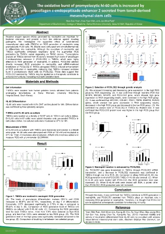
     Cell information                                          Figure 2. Selection of PCOLCE2 through genetic analysis
     T-MSCs were isolated from human palatine tonsils obtained from patients  (A). We analyzed increasing and decreasing gene expression in the high ROS
     undergoing  tonsillectomy  at  Ewha  Womans  University  Mok-Dong  group by RNA sequencing. (B). It was confirmed through real-time PCR that
     Hospital(Seoul, South Korea).                             SFRP4, MEDAG, MFAP5, and PCOLCE2 gene, which showed high gene
                                                               expression in RNA sequencing results, were increased in the high ROS group.
                                                               (C). It was confirmed that APCDD1, COL8A1, PSG1, PDPN, SNAP25, RUNX3
     HL-60 Differentiation                                     genes, which showed low gene expression in RNA sequencing results,
     HL-60 cells were treated with 0.8% DMF and incubated for 96h. Differentiation  decreased in the high ROS group and increased in the low ROS group. (D). We
     was confirmed by flow cytometry analysis.                 confirmed the protein level of PCOLCE2 in TMSCs by Western blot. It was
                                                               confirmed that PCOLCE2 protein level was higher in the high ROS group than
     T-MSCs and HL-60 cells co-culture                         low ROS group.
     TMSCs were seeded at a density of 5x10 cells in 100mm cell culture dishes.  A                                                                              B
                                 5
                  6
     D-HL-60 cells(1x10 cells) were added dropwise onto pre-seeded TMSCs in
     cell culture dishes. Co-cultured cells were incubated at 37℃ for 48h.  1 5 0                 C o n tro l  P M A
                                                                            ****                4 0 0  * * * *  * * *  *
                                                                    R e la tiv e  P C O L C E 2  e x p re s s io n  (fo ld )  1 0 0  3 0 0
     Measurement of ROS                                               5 0                       R O S  g e n e ra tio n (%  o f P M A  tre a te d  D -H L -6 0 )  2 0 0
     D-HL-60 cells co-cultured with TMSCs were harvested and seeded in a 96well                 1 0 0
     white plate. HL-60 cells were stimulated with PMA of 100 nM and incubated at  0             0
     37℃ for 15min. Immediately after incubation, 300μM of luminol was added and  s iN e g -T M S C  s iP C O L C E 2 -T M S C  H L -6 0  D -H L -6 0
                                                                                                      s iP C O L C E 2 -T M S C + D -H L -6 0
     measure ROS with a luminometer GLOWMAX.                                                        s iN e g -T M S C + D -H L -6 0
                            Result                                C                                                                               D
                                                                       C o n tro l  P M A         C o n tro l  P M A
    A                                                                                         B                                    * * * *
                                                                             * *                 2 5 0  * * *
             PE     FITC   APC                                        3 0 0  *                       * * * *
                                          C o n tro l  P M A          2 0 0  *                   2 0 0  * * * *
                                                                                                 1 5 0
       HL-60                            2 0 0  * * *  * * *  * * *  R O S  g e n e ra tio n (%  o f P M A  tre a te d  D -H L -6 0 )  1 0 0  R a te  o f R O S  g e n e ra tio n (%  o f P M A  tre a te d  D -H L -6 0 )  1 0 0
                                       R O S  g e n e ra tio n (%  o f P M A  tre a te d  D -H L -6 0 )  1 0 0  0  0  0 .1  1  2  1 0 0  3 0 0  5 0 0  0  1 0 0  5 0 0  1 0 0  5 0 0  (n g /m l)
                                        1 5 0
                                                                                                     P C O L C E 2
                                                                          P C O L C E 2  (n g /m l)
      dHL-60                            5 0 0                  Figure 3. Neutrophil function is enhanced by PCOLCE2.  B S A
                                          H L -6 0  D -H L -6 0  T M S C    B M S C  (A). PCOLCE2 was gene knockdown in TMSCs through PCOLCE2 siRNA
                                                               transfection, and a decrease in PCOLCE2 expression was confirmed in
    C                                                                                       D                                 E                              TMSCs through real time PCR. (B). Compared to siNeg-TMSC+D-HL-60, the
                                                               ROS generation level of siPCOLCE2-TMSC+D-HL-60 was lowered. (C). When
      3 0 0
     R O S  g e n e ra tio n (%  o f P M A  tre a te d  D -H L -6 0 )  2 0 0 1 0 0  170%  R O S  g e n e ra tio n (%  o f P M A  tre a te d  D -H L -6 0 )  2 5 0 2 0 0 1 5 0  * * * *  D o u b lin g  tim e  (h )  5 0 4 0 3 0  D-HL-60 cells were treated with PCOLCE2, the ROS generation level was
                                                               increased. (D). When D-HL-60 cells were treated with BSA, a protein other
                                  120%
                                                               than PCOLCE2, ROS production was not increased.
       0
       T M S C 6 T M S C 3 T M S C 9 T M S C 2 T M S C 4 T M S C 1 7 T M S C 7 T M S C 2 1 T M S C 1 0 T M S C 1 3 T M S C 8 T M S C 1 1 T M S C 1 5 T M S C 1 8 T M S C 2 4 T M S C 1 2 T M S C 2 3 T M S C 1 T M S C 2 5 T M S C 1 6 T M S C 1 4 T M S C 5 T M S C 1 9 T M S C 2 2 T M S C 2 0  1 0 0 5 0 0  2 0 1 0 0  Conclusion
        H ig h  R O S       L o w  R O S  H ig h  R O S  L o w  R O S  H ig h  R O S  L o w  R O S
                                                               Through this study, it was confirmed that TMSCs contribute to ROS generation in
     Figure 1. TMSCs are involved in neutrophil ROS generation.  neutrophils, and PCOLCE2 secreted from TMSCs plays an important role in
     (A). The levels of granulocyte differentiation markers CD11b and CD35  increasing ROS generation in neutrophils. Therefore, it is thought that PCOLCE2
     increased to 98.97% and 82.16%, respectively, on day 4 of differentiation.  can be applied as a therapeutic candidate for enhancing immunity.
     Conversely, CD71 decreased significantly to 2.76% in day 4 compared to
     98.25% in day 0. (B). Comparison of ROS generation level of TMSCs and          Reference
     BMSCs co-cultured with D-HL-60 cells. (C). Comparing the ROS generation
     level for each TMSCs donor, more than 170% were selected as high ROS  Yoon Shin Park, Goh-Woon Lim, Kyung-Ah Cho, So-Youn Woo, Mee young Shin,
     group, and less than 120% were selected as low ROS group. (D). The ROS  Eun-Sun Yoo, Jeong Chan Ra, Kyung-Ha Ryu, (2012) Improved viability and
     generation level of the high group was significantly increased compared to the  activity of neutrophils differentiated from HL-60 cells by co-culture with adipose
     low group. (E). Comparison of doubling time between two selected groups.  tissue-derived mesenchymal stem cells, Biochem Biophys Res Commun, Jun
                                                               22;423(1):19-25

     RESEARCH POSTER PRESENTATION DESIGN ©  2019
     www.PosterPresentations.com

