
Page 19 - U. Protein structure and function
P. 19
The Dynamic Structure of human Interleukin 33 in the apo form
Se-Young Son , Sang-Hyun Son , Young-Ha Ryu and Young Ho Jeon 1,2
1
1
1
1 Allercuris Co. Ltd., Lab.402, College of Pharmacy, Sejong-ro 2511, Sejong 30019, Korea,
2 College of Pharmacy, Korea University, Sejong-ro 2511, Sejong 30019, Korea
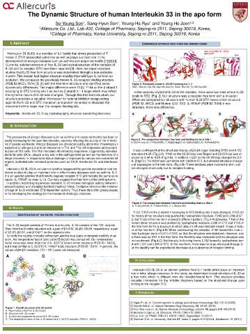
I. ABSTRACT COMPARISON BETWEEN FREE FORM AND BINDING FORMS
(red) IL-33 (PDB ID : 4KC3)
Interleukin 33 (IL33) is a member of IL1 family that drives production of T Site2 (plum) ST2
helper-2 (Th2) associated cytokines as well as plays a critical role in the Site1
development of allergic diseases such as asthma and atopic dermatitis [1] [2] [3].
Currently, solution structure of free IL-33 and crystal structure of the complex of
IL-33 and its receptor ST2 have been reported [4]. Here, we report structure
studies that IL-33 free form structure was determined through a non-oxidative
mutant. This mutant had higher structure stability than wild type IL-33 from an
oxidation. We compared the previously known IL-33-receptor binding structure
(PDB ID:4KC3, 5VI4) [5] [6] with the free form structure and identified some Figure 2. Previously known IL-33:ST2 binding site 1, 2
dynamically differences. The major difference were Y122, Y164 on the β strand 1 Site1: Glu144, Glu148, Asp149, Asp144, Site2: Glu165, Tyr163, Leu182
belonging to ST2 binding site 2 as well as β strand 3 ~ 4 loops which may reflect In the complex crystal of IL-33 to the receptor, there were two sites where IL-33
the dynamic nature of IL-33 was changed. Through this free form structure, our binds to ST2. (Fig. 2) Our structure was a receptor-free form with a mutation.
structure provides a valuable information for rational inhibitor design acting When we compared our structure with human IL33:ST2 hetero dimer structure
against the IL-33 and ST2 interaction and protein dynamics to elucidate the (PDB ID: 4KC3) and Murine IL33: ST2: IL-1RAcP (PDB ID: 5VI4) trimer
movement of the loops near the receptor binding site. structure, there was difference.
Keywords : Interleukin 33, X-ray crystallography, structure-based drug discovery A B C D
II. INTRODUCTION
The prevalence of allergic diseases such as asthma and atopic dermatitis has been st
eadily increasing for the past few decades, severely affecting the quality of life of millio Figure 3. The comparison between free form and binding forms at Site 1
A. Β3-β4 loop, B. Y143(mY140), C. N222(mL221) & H224(mS223), D. H246(mQ245)
ns of people worldwide. Allergic diseases are characterized by abnormal inflammatory r
esponses to allergens due to an imbalance in Th1 and Th2 cell responses and overpro
duction of Th2 cells. The Th2-mediated inflammatory responses, triggered by activated It was confirmed that the structural change of β3-β4 loops including D153 and K152
dendritic cells (DCs), played a key role in the pathogenesis as well as maintenance of a was about 4.9Å (Fig.3-A). Y143 on the β2-β3 loop was flipped and β2-β3 loop was ch
llergic diseases. In response to tissue damage or exposure to various environmental all anged by 3.4Å to 4.8Å (FIg.3-B). In addition, H224 on the β8-β9 loop changed by 2.9
ergens, epithelial cells released cytokines such as TSLP, Interleukin 33, and Interleukin Å. (Fig3-C) The N222 was combined with H309 of ST2, but showed structural change
25. s of asparagine in the free form. (Fig.3-B) These residues were involved in site1 and
Interleukin 33 (IL-33) is an alarmin cytokine suggested by genetic association and fun are changed structurally due to receptor binding.
ctional studies to play an important role in inflammatory diseases such as asthma. IL-3
3 is an agonist cytokine that binds its cognate receptor ST2 and recruits the same co re A B C
ceptor, IL-1RAcP, as does IL-1b. Our data suggest that free form unlike binding forms.
In addition, According to previous research, IL-33 showed biological activity between r
educed (active) and disulpide bonded (inactive) forms. Oxidation-driven conformational
change of IL-33 eliminates ST2-dependent activity. Thus these data offer great prospec
ts for developing the strategy for the treatment of allergic diseases.
Figure 4. The comparison between free form and binding forms at Site 2
A. Y122(mS119), B. Y163(mS160), C. L182(mL180)
III. RESULTS
Y122, Y163 on the β strand 1 belonging to ST2 binding site 2 was changed. Y122 of t
IDENTIFICATION OF THE ENTIRE PROTEIN STRUCTURE he hetero dimer structure was pushed by hydrophobic residues, F245 and L246 of ST
2, but Y122 of the free form showed a different pattern. (Fig.4-A) Likewise, Y163 of the
hetero dimer structure was pushed by hydrophobic residues, L308 of ST2, but Y122 of
The IL-33 mutant consists of 11mers in one units. IL-33 consists of the 12β -strands. the free form forms a structure in which about 2.3Å was directed inward. It was a featu
They form the β-trefoil structure with a pair of β1-β12, β4-β5, β8-β9, respectively, a pair re of the free form. (Fig.4-B) When constructing the complex, K180 formed the main c
of β2-β3, β6-β7, and β10-β11 on the opposite side. hain hydrogen bond of L311 of ST2, so that the structure was stabilized. However, sinc
To verify the validity of model refinement and the true static or dynamic mobility of an e there was no ST2 in the free form, the flexibility was increased and the structure was
atom, the temperature factor (also called B factor) was compared. Our temperature not confirmed. (Fig.4-C) Additionally, In binding forms, L182 formed a hydrophobic bon
factor value was lower than that of IL-33:ST2 hetero dimer structure (PDB ID : 4KC3), d with L311 and L306 of ST2. In the free form, there was no large structural change, b
but It was similar to IL-33:ST2:IL-1RAcP triple structure (PDB ID : 5VI4). In general, the ut the stability can be expected to decrease due to absence of receptor binding.
values of β4~β5 (residues 170 ~ 181) were not measured.
A C D
IV. CONCLUSION
Interleukin-33 (IL-33) is an alarmin cytokine from IL-1 family which plays an important
role in initial allergic response. In this study, we determined crystal structure of IL-33 as
mutant
a free form, which is different from the receptor-bound form. This structure provides
valuable information for the inhibitor discovery based on the structural change upon
B binding to the receptor ST2.
V. REFERENCES
[1] Ngoc PL et. ol. Current opinion in allergy and clinical immunology. 5(2):161-6 (2005)
[2] Gandhi NA et. ol. Nature Reviews Drug Discovery, 15, 35-50 (2016)
Figure 1. Overall structure of IL-33 mutant [3] Foo Yew Liew et. ol. Nature Reviews Immunology, volume 16, pages 676–89 (2016)
A : Asymmetric unit of the IL-33 mutant
B : Structure of IL-33 mutant [4] Andreas Lingel et. ol. Structure, 17(10):1398-410 (2009)
C : β-trefoil and supposed structure of IL-33 mutant [5] Liu, X et. ol. PNAS, 110 (37) 14918-14923, September 10 (2013)
D : The temperature factor of the IL-33 mutant and IL-33:ST2 complex (PDB ID : 4KC3) [6] Günther S et. ol. Immunity, Volume 47, Issue 3, pages 510-523.e4, September 19 (2017)