
Page 7 - T. Protein modification and regulation
P. 7
Regulation of Fra-1 protein stability through deubiquitination promotes colorectal
cancer metastasis
Sun-Il Yun 1 , Hye Kyung Hong 2 , So-Young Yeo 3 , Seok-Hyung Kim 3,4* , Yong Beom Cho 2,4* , and Kyeong Kyu Kim 1,4*
1Department of Precision Medicine, Sungkyunkwan University School of Medicine, Suwon, 2Department of Surgery, Samsung Medical Center, Sungkyunkwan University School of Medicine, Seoul, 3Department of Pathology, Samsung Medical Center, Sungkyunkwan University School of Medicine, Seoul, 4Samsung Medical
Center, Department of Health Science and Technology, Samsung Advanced Institute for Health Science and Technology, Sungkyunkwan University School of Medicine, Seoul.
BACKGROUND AIM
Due to the advances in screening techniques and targeted cancer therapy, the death rate from colorectal cancer The ubiquitin-dependent protein stability of Fra-1 has not been as well studied as that of c-Jun, the
(CRC) has been decreasing. However, CRC is the second leading cause of cancer death in the world, with a poor representative heterodimeric partner of Fra-1 (Vial and Marshall., 2003). Therefore, we screened for
five-year survival for patients diagnosed with metastatic colorectal cancer (mCRC) (Engstrand et al., 2018). Therefore, deubiquitinase enzymes (DUBs) that show strong deubiquitination activity in 293T cells by monitoring the
a targeted therapy for mCRC is necessary for effective treatment of CRC to improve survival rate. amount of total ubiquitinated protein after transfection with DUB-encoding plasmids (Yun et al., 2015).
Activator protein 1 (AP-1) is one of transcription factors and composes of members of Fos (c-Fos, FosB, Fra-1, and
Fra-2) and Jun (c-Jun, JunB, and JunD) family. Fos proteins can only heterodimerize with Jun proteins which can Among the tested DUBs, we tried to newly identify a Fra-1 DUB in CRC and to investigate the oncogenic role
homodimerize and heterodimerize with Fos members. The Fos protein is intrinsically unstable and stabilized by ERK of DUB in cancer metastasis related with Fra-1 expression.
activation and the Fra-1 is an activator of transcriptional activity by binding to AP-1 site and this is also regulated by
MAPK pathway.
METHODS
Cell culture : 293T and HCT116 cells were cultured in DMEM and HT-29 cells in RPMI1640 medium supplemented with 10% heat-inactivated FBS containing penicillin-streptomycin at 37°C in the presence of 5% CO 2 .
Plasmids: Full-length human USP21 (NM_001014443.2) was cloned into pDEST-CMV6 with the SRT tag at the N-terminus. The catalytic residue mutant USP21 C221A was generated using PCR mutation.
USP21 knockdown: the following USP21 siRNA sequence (NM_001014443.2) was purchased from Bioneer.
Immunoblotting and immunoprecipitation (IP): Cells were solubilized in RIPA lysis buffer then enriched and detected with indicated antibodies.
Invasion assay and Migration assay: Both assays were performed in a 24-well invasion chamber with an 8 m pore size polycarbonate membrane according to the manufacturer’s instruction (Millipore).
Collection and tissue microarray (TMA) of human CRC samples: Tissue specimens from the tumors of 374 patients who had undergone curative surgery for colorectal cancer at Samsung Medical Center, Sungkyunkwan
University School of Medicine, Seoul, South Korea were used. TMA was constructed using a manual tissue arrayer (Beecher Instruments).
Animals and splenectomy: BALB/c nu/nu mice (6-8 weeks old, female, n=6 per group), A small left abdominal flank incision was made, and the spleen was exteriorized for intra-splenic injection, then prepared cells were injected
into the spleen. After splenic vein was surtured, the spleen was dissected.
In vivo tumor imaging: After cancer cell injection, MRI imaging (Biospec 7T, Bruker) was initiated at Day 14 and repeated once a week for as long as liver metastasis was detected.
Immunohistochemistry (IHC): Sections were stained with hematoxylin and eosin (H&E) to examine tumor morphology then, USP21 and Fra-1 were detected.
IHC and evaluation of CRC samples: Sections of the TMA were immunohistochemically labeled with USP21 using a BenchMark XT automated stainer (Ventana Medical Systems, Inc.), according to the manufacturer’s protocol.
Then, to evaluate expression of USP21, an IHC score was generated by multiplying the percentage of USP21-positive cells by staining intensity.
Statistics: Statistical comparisons between groups were conducted using paired t-test or analysis of variance (ANOVA) and CRC samples Statistical analyses were performed using SPSS 18.0 statistical software (SPSS). Chi-
square tests (Pearson’s chi-square test or chi-square test using linear by linear association) were used to analyze correlations between IHC results and clinicopathologic parameters. Survival curves were plotted using the Kaplan–
Meier method.
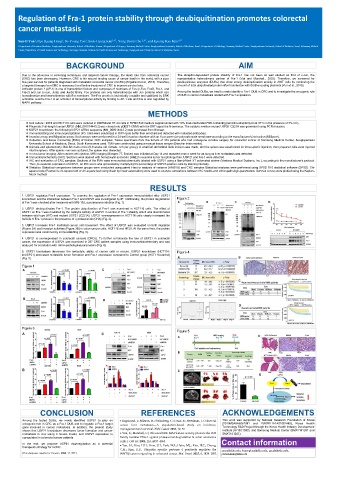
RESULTS
1. USP21 regulates Fra-1 expression. To examine the regulation of Fra-1 expression immunoblotted after USP21
knockdown and the interaction between Fra-1 and USP21 was investigated by IP. Additionally, the protein degradation Figure 2
of Fra-1 was checked after treatment with MG-132, a proteasome inhibitor (Fig. 1)
2. USP21 deubiquitinates Fra-1. The protein ubiquitination of Fra-1 was examined in HCT116 cells. The effect of
USP21 on Fra-1 was mediated by the catalytic activity of USP21 in control of Fra-1 stability which was discriminated
between wild type (WT) and mutant USP21 (C221A). USP21 overexpression in HCT116 cells clearly increased the
half-life of Fra-1 protein in the presence of cycloheximide (CHX) (Fig. 2).
3. USP21 increases Fra-1 mediated cancer cell movement. The effect of USP21 was evaluated on both migration
(Figure 3A) and invasion activities (Figure 3B) in colon cancer cells, HCT116 and HT29. At the same time, the protein
expression was examined by immunoblotting (Fig. 3).
4. USP21 is overexpressed in colorectal cancers (CRCs). To further corroborate the role of USP21 in colorectal
cancer, the expression of USP21 was examined in 297 CRC patient samples using immunohistochemistry and was
analyzed for correlation with clinicopathological parameters (Fig. 4).
5. USP21 knockdown decreases the metastatic ability of cancer cells in mouse. USP21 knockdown (HCT116- Figure 4
shUSP21) decreased metastatic tumor formation and Fra-1 expression compared to Control group (HCT116-control)
(Fig. 5).
Figure 1
Figure 3
Figure 5
CONCLUSION REFERENCES ACKNOWLEDGEMENTS
Among the tested DUBs, we newly identified USP21 to play an Engstrand, J.; Nilsson, H.; Strömberg, C.; Jonas, E.; Freedman, J. Colorectal This work was supported by National Research Foundation of Korea
oncogenic role in CRC as a Fra-1 DUB and to regulate a Fra-1 target cancer liver metastases—A population-based study on incidence, (2019M3A9A8067081 and 2020R1I1A1A01052480), Korea Health
gene involved in cancer metastasis. In addition, the present study management and survival. BMC Cancer 2018, 18, 78 Technology R&D Project through the Korea Health Industry Development
shows that USP21 knockdown decreases tumor formation and cancer Vial, E.; Marshall, C.J. Elevated ERK-MAP kinase activity protects the FOS Institute (HI15C1593), and Samsung Medical Center (SMX1161261 and
metastasis in vivo using a mouse model, and USP21 expression is SMX1161241).
upregulated in colorectal cancer patients. family member FRA-1 against proteasomal degradation in colon carcinoma
cells. J. Cell Sci. 2003, 116, 4957–4963.
In the end, we propose USP21 downregulation as a potential Yun, S.I.; Kim, H.H.; Yoon, J.H.; Park, W.S.; Hahn, M.J.; Kim, H.C.; Chung, Contact information
therapeutic strategy for mCRC.
C.H.; Kim, K.K. Ubiquitin specific protease 4 positively regulates the yunsi@skku.edu, kyeongkyu@skku.edu, gscyb@skku.edu,
(This study was reported in ‘Cancers, 2020, 12, 207’.) WNT/β-catenin signaling in colorectal cancer. Mol. Oncol. 2015, 9, 1834–1851. platoshkim@skku.edu