
Page 11 - T. Protein modification and regulation
P. 11
.
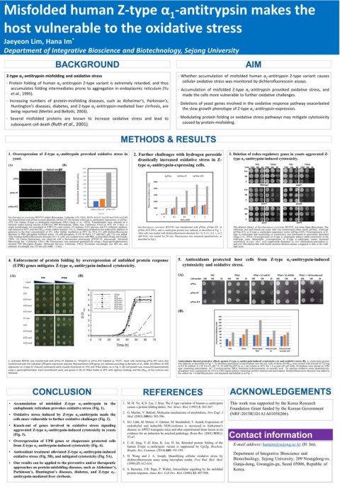
Misfolded human Z-type α -antitrypsin makes the
1
host vulnerable to the oxidative stress
Jaeyeon Lim, Hana Im *
Department of Integrative Bioscience and Biotechnology, Sejong University
BACKGROUND AIM
Z-type α 1 -antitrypsin misfolding and oxidative stress · Whether accumulation of misfolded human α 1 -antitrypsin Z-type variant causes
· Protein folding of human α 1 -antitrypsin Z-type variant is extremely retarded, and thus cellular oxidative stress was monitored by dichlorofluorescein assays.
accumulates folding intermediates prone to aggregation in endoplasmic reticulum (Yu · Accumulation of misfolded Z-type α 1 -antitrypsin provoked oxidative stress, and
et al., 1995). made the cells more vulnerable to further oxidative challenges.
· Increasing numbers of protein-misfolding diseases, such as Alzheimer’s, Parkinson’s, · Deletions of yeast genes involved in the oxidative response pathway exacerbated
Huntington’s diseases, diabetes, and Z-type α 1 -antitrypsin-mediated liver cirrhosis, are the slow growth phenotype of Z-type α 1 -antitrypsin-expression.
being reported (Merlini and Bellotti, 2003).
· Several misfolded proteins are known to increase oxidative stress and lead to · Modulating protein folding or oxidative stress pathways may mitigate cytotoxicity
subsequent cell death (Ruth et al., 2001). caused by protein-misfolding.
METHODS & RESULTS
1. Overexpression of Z-type α 1 -antitrypsin provoked oxidative stress in 2. Further challenges with hydrogen peroxide 3. Deletion of redox-regulatory genes in yeasts aggravated Z-
yeast. drastically increased oxidative stress in Z- type α 1 -antitrypsin-induced cytotoxicity.
(A) (B) type α 1 -antitrypsin-expressing cells. YPD YPGal YPD YPGal
Dichlorofluorescein Optical merge 1000 Cells 1500 300 60 1500 300 60 1500 300 60 1500 300 60
900 Vec
Fluorescence (Arbitrary units) 500 4000 Vec Wild-type ∆yap1
pYInu 800 5000 pYInu pYInu-AT pYInu-ATZ Wt Z
700
600
pYInu-AT 400 Fluorescence (Arbitrary Units) 3000 Wt
300
200
pYInu-ATZ 100 0 2000 Z ∆skn7 ∆sod2
1000
pYInu pYInu-AT pYInu-ATZ Vec
Strains 0 Wt
Saccharomyces cerevisiae BY4741 (Open Biosystems, Lafayette, CO, USA; MATa his3Δ1 leu2Δ0 met15Δ0 ura3Δ0) 0 0.1 0.5 1 2
was transformed with pYInu (a mock plasmid), pYInu-AT (for human wild-type α 1 -antitrypsin expression), or pYInu- H 2 O 2 (mM) Z
ATZ (for human Z-type α 1 -antitrypsin expression) DNA (Jung et al., 2014). Transformants were selected on a ∆tsa1 ∆pst2
minimal media lacking leucine (CSM-Ura; MP Biomedicals, Santa Ana, California, USA) at 30°C for 3 days. A
single transformant was inoculated in YPR (1% yeast extract, 2% peptone, 0.2% glucose, and 2% raffinose) medium, Saccharomyces cerevisiae BY4741 was transformed with pYInu, pYInu-AT, or The deletion library of Saccharomyces cerevisiae BY4741 was from Open Biosystems. The
and cultured at 30°C until the OD 600 of the culture reached ~0.4. α 1 -Antitrypsin production was induced by addition of pYInu-ATZ DNA, and α 1 -antitrypsin protein was induced, as described in Fig. 1. wild-type and each knock-out yeast cells was transformed either mock (pYInu), wild-type
galactose to the final concentration of 2%. After further incubation at 30°C for 20 h, 0.1 mM dichlorofluorescein After cells was loaded with dichlorofluorescein diacetate for 1 h, 0, 0.1, 0.5, 1, or 2 (pYInu-AT) or Z-type α 1 -antitrypsin expression vector (pYInu-ATZ). Overexpression of Z-
diacetate in PBS (phosphate buffered saline; 10 mM phosphate, 0.138 M NaCl, 2.7 mM KCl, pH 7.4) was added. type α 1 -antitrypsin and monitoring of cytotoxicity was performed as previously described
After loading dichlorofluorescein diacetate for 1 h, cells was washed twice with PBS, as described (Wang and Joseph, mM H 2 O 2 was treated for 20 min. Fluorescence was measured quantitatively, as (Jung et al., 2014). Overexpression of Z-type α 1 -antitrypsin induces mild cytotoxicity in the
1999). (A) Green fluorescence was observed with a fluorescence microscopy (EVOS FL microscope, Advanced described in Fig.1. wild-type yeast. Meanwhile, overexpression of Z-type α 1 -antitrypsin causes increased
Microscopy Inc., California, USA). (B) Fluorescence was measured quantitatively using a fluorespectrophotometer cytotoxicity in yap1, skn7, sod2 (superoxide dismutase 2), tsa1 (thioredoxin peroxidase 1),
(Gemini EM Microplate Reader, Molecular Devices, California, USA). Excitation wavelength was 485 nm, and and pst2 (flavodoxin-like fold family protein) deletion strains, compared to that in the wild-
emission wavelength was 535 nm (slit width = 5 nm). type yeast.
4. Enforcement of protein folding by overexpression of unfolded protein response 5. Antioxidants protected host cells from Z-type α 1 -antitrypsin-induced
(UPR) genes mitigates Z-type α 1 -antitrypsin-induced cytotoxicity. cytotoxicity and oxidative stress.
(A) (B) (A) YPD YPGal YPGal + 0.5 mM AC YPGal + 10 µM BHA YPGal + 1 mM Ascorbic acid
YPD YPGal
Cell number 500 100 20 500 100 20 500 100 20 500 100 20 500 100 20
Cell #
pYInu
AT(wt)
9
pYInu-AT
ATZ 8 AT(wt)
ATZ
ATZ+lhs1 7 ATZ+lhs1
ATZ+hac1
ATZ+ent1 pYInu-ATZ
ATZ+hac1 6 ATZ+kar2
ATZ+ssa1
O.D. 600 4
ATZ+ent1 5 ATZ+ssa2 (B)
AT (wt) 200
3 180
160
ATZ 2 140
ATZ+kar2 1 Fluorescence (Arbitrary units) 120
100
0 80
ATZ+ssa1 60
0 10 20 30 40 50 60 70 40
ATZ+ssa2 20
Time (h) 0
YPD YPGal YPGal+10μM YPGal+0.5mM YPGal+0.5mM
BHA AC Ascorbic acid
S. cerevisiae BY4741 was transformed with pYInu-AT [labeled as “AT(wt)”] or pYInu-ATZ (labeled as “ATZ”). Yeast cells harboring pYInu-ATZ were also Antioxidants showed protective effects against Z-type α 1 -antitrypsin-induced cytotoxicity (A) and oxidative stress (B). α 1 -Antitrypsin protein
transformed with the indicated UPR gene expression plasmid. Representative UPR genes are selected according to Bernales et al., 2006. (A) Effects of UPR was induced, as described in Fig. 1. To facilitate penetration of antioxidants into the cell, cells of about 20 OD 600 were treated with lyticase (0.725 mg
expression on Z-type AT-induced cytotoxicity were visually monitored on YPD and YPGal plates, as in Fig. 3. (B) Cell growth was measured quantitatively in 0.9 M sorbitol, 0.1 M Tris-Cl, pH 7.5, 25 mM Na 2 EDTA) in 1 ml volume at 30°C for 1 h to peel off cell walls. Protoplasts were spread on YPG
using a spectrophotometer. Each recombinant yeast was grown in 30 ml YPGal media at 30°C with vigorous shaking, and the OD 600 of the cultures was agar containing antioxidants: AC, N-acetylcysteine; BHA, butylated hydroxyaninole; or ascorbic acid. Το monitor oxidative stress quantitatively,
protoplasts were regenerated for 24 h in YPG liquid cultures containing sorbitol solution and antioxidants. Dichlorofluorescein diacetate was added to
followed. the culture for 1 h and fluorescence was measured and labeled as in Fig. 1.
CONCLUSION REFERENCES ACKNOWLEDGEMENTS
• Accumulation of misfolded Z-type α 1 -antitrypsin in the 1. M.-H. Yu, K.N. Lee, J. Kim, The Z type variation of human α 1 -antitrypsin This work was supported by the Korea Research
endoplasmic reticulum provokes oxidative stress (Fig. 1). causes a protein folding defect, Nat. Struct. Biol. (1995) 2: 363-367. Foundation Grant funded by the Korean Government
• Oxidative stress induced by Z-type α 1 -antitrypsin made the 2. G. Merlini, V. Bellotti, Molecular mechanisms of amyloidosis, New Engl. J. (NRF-2015R1D1A1A01058206).
Med. (2003) 349(6): 583-596.
cells more vulnerable to further oxidative challenges (Fig. 2).
3. H.J. Lüth, M. Holzer, U. Gärtner, M. Staufenbiel, T. Arendt, Expression of
• Knock-out of genes involved in oxidative stress signaling endothelial and inducible NOS-isoforms is increased in Alzheimer's
aggravated Z-type α 1 -antitrypsin-induced cytotoxicity in yeasts disease, in APP23 transgenic mice and after experimental brain lesion in rat:
(Fig. 3). evidence for an induction by amyloid pathology, Brain Res. (2001) 913(1): Contact information
57-67.
• Overexpression of UPR genes or chaperones protected cells 4. C.-H. Jung, Y.-H. Kim, K. Lee, H. Im, Retarded protein folding of the
from Z-type α 1 -antitrypsin-induced cytotoxicity (Fig. 4). human Z-type α 1 -antitrypsin variant is suppressed by Cpr2p, Biochem. E-mail address: hanaim@sejong.ac.kr (H. Im).
• Antioxidant treatment alleviated Z-type α 1 -antitrypsin-induced Biophy. Res. Commun. (2014) 445: 191-195. Department of Integrative Bioscience and
oxidative stress (Fig. 5B), and mitigated cytotoxicity (Fig. 5A). 5. H. Wang and J. A. Joseph, Quantifying cellular oxidative stress by
dichlorofluorescein assay using microplate reader, Free Rad. Biol. Med. Biotechnology, Sejong University, 209 Neungdong-ro,
• Our results can be applied to the preventive and/or therapeutic (1999) 27: 612-616. Gunja-dong, Gwangjin-gu, Seoul 05006, Republic of
approaches on protein-misfolding diseases, such as Alzheimer’s, 6. S. Bernales, F.R. Papa, P. Walter, Intracellular signaling by the unfolded Korea.
Parkinson’s, Huntington’s diseases, diabetes, and Z-type α 1 - protein response, Annu. Rev. Cell Dev. Biol. (2006) 22: 487-508.
antitrypsin-mediated liver cirrhosis.