
Page 1 - R. Organoid
P. 1
Characterization of iPSC-derived midbrain organoids from Parkinson’s disease patient
Yunsu Bang, Juhyun Choi, Ki Soon Kim, Jong Gu Lee, Woo yong Oh, Yoonsook Lee
Clinical Research Division, NIFDS, MFDS, Osong Health Technology Administration Complex, 187, Osongsaengmyeong 2-ro, Osong, Heungdeok, Cheongju, 28159, Korea
Abstract
Parkinson's disease(PD) is the one of common neurodegenerative diseases, that results from the loss of neuromelanin(NM)-containing dopaminergic(DA) neurons. To overcome the
limitations of animal degenerative models for human phenotypes, human induced Pluripotent Stem Cell(iPSCs) are being widely used nowadays. In this study, we investigated the
characterization of midbrain organoids(MOs) whether these can be the proper model for research and drug screening on neurodegenerative diseases.
At first, we differentiated MOs from human iPSCs by 3D culture system. And then we checked the identified markers for representing DA neurons on culture day 0, 24, 44, 64, 84 by
real time PCR, WB, and IHC. The differentiated MOs showed that DA progenitors(FoxA2, LMX1A and LMX1B), neuronal microtubule(Tuj1, MAP2) and DA neuron(TH, GIRK2)
markers including accumulation of alpha-synuclein. First of all, the NM granules were observed in organoids, not shown in 2D-cultured iPSCs. NM granules of patient's MOs were
detected lower than those of normals', that shows the major characteristic of PD.
Our study showed that the MOs had several features of the human midbrain. Even though more assessments remain to be elucidated, we propose that iPSCs-derived organoids are
useful tool for researches.
Material and Methods
Analysis of differentiation
To confirm the expression of differentiation markers, such as Oct4, NANOG, LMX1A, TH and Tuj1, Real-Time PCR, Western blotting and Immunohistochemistry were performed.
The PCR test was performed following the ssoadvanced universal probes supermix(Bio-Rad) protocol and used following the primers, GAPDH, OCT4, NANOG, LMX1A, TH and
Tuj1(Applied biosystems). The expression of proteins was assessed with antibodies such as OCT4(Cell Signaling Technology), LMX1B(Abcam), TH(Abcam), α-synuclein(Abcam),
Dopamine(Abcam) and actin(Sigma) by western blot analysis and immunohistochemistry.
L-DOPA treatment
L-DOPA is metabolized to dopamine in dopaminergic neurons. We treated the cells with excess L-DOPA and observed the remaining dopamine in the cells accumulated as
neuromelanin. L-DOPA is diluted with organoid culture medium to a final concentration of 50 μM for more than 1 month.
Results
Differentiation of human midbrain organoids
Midbrain organoid differentiation was induced using modifications to the Junghyun Jo’s
protocol as below(Figure 1). Induced Pluripotent Stem Cells(iPSCs) were obtained from
normal and Parkinson’s disease patient.
A.
B.
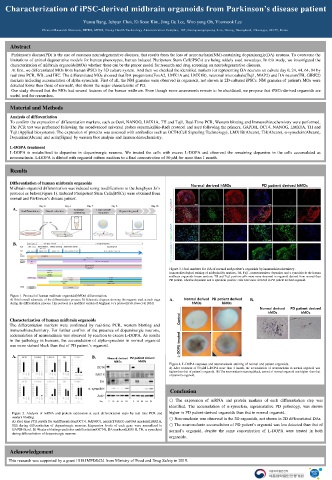
Figure 3. Final markers for DA of normal and patient’s organoids by immunohistochemistry .
Immunohistological staining of midbrain DA markers, TH, Tuj1, neurotransmitter dopamine and α-synculein in the human
midbrain organoids frozen sections. TH and Tuj1 positive cells were more observed in organoid derived from normal than
PD patient, whereas dopamine and α-synculein positive cells were more detected in PD patient derived organoid.
Figure 1. Protocol of human midbrain organoids(hMOs) differentiation.
A) Brief overall schematic of the differentiation process; B) Schematic diagram showing the reagents used at each stage A. B.
during the differentiation process. This protocol is a modified version of Junghyun Jo’s protocol(Cell Stem Cell, 2016)
Characterization of human midbrain organoids
The differentiation markers were confirmed by real-time PCR, western blotting and
immunohistochemistry. For further confirm of the presence of dopaminergic neurons,
accumulation of neuromelanin was observed by reaction to excess L-DOPA. As results
in the pathology in humans, the accumulation of alpha-synuclein in normal organoid
was more stained black than that of PD patient’s organoid.
A. B.
Figure 4. L-DOPA response and neuromelanin staining of normal and patient organoids.
A) After treatment of 50 μM L-DOPA more than 1 month, the accumulation of neuromelanin in normal organoid was
higher than that of patient’s organoid. B) The neuromelanin staining(black, arrow) of normal organoid was higher than that
of patient’s organoid.
Conclusion
○ The expression of mRNA and protein markers of each differentiation step was
identified. The accumulation of α-synuclein, representative PD pathology, was shown
Figure 2. Analysis of mRNA and protein expression at each differentiation steps by real time PCR and higher in PD patient-derived organoids than that in normal organoid.
western blotting. ○ Neuromelanin was observed in the 3D organoids, not shown in 2D differentiated DAs.
A) Real time PCR results for undifferentiation(OCT4, NANOG), neural(TUBB3) and DA markers(LMX1A,
TH) during differentiation of dopaminergic neurons. Expression levels of each gene were normalized to ○ The neuromelanin accumulation of PD patient’s organoid was less detected than that of
GAPDH level. B) Western blotting results for undiffentiation(OCT4), DA markers(LMX1B, TH, α-synuclein) normal’s organoid, despite the same concentration of L-DOPA were treated in both
during differentiation of dopaminergic neurons.
organoids.
Acknowledgement
This research was supported by a grant 19181MFDS424 from Ministry of Food and Drug Safety in 2019.