
Page 75 - Q. Neuroscience
P. 75
The dopaminergic circuit in control of compulsive eating behavior
Bokyeong Kim and Ja-Hyun Baik
Molecular Neurobiology Laboratory, Department of Life Sciences, Korea University, Seoul 02841, Republic of Korea
BACKGROUND AIM
Dopamine serves a central role in motivated behavior and reward processing, in which Palatable food drives hedonic food consumption, and hedonic drive to feed is a key contributor to food
dopamine D2 receptor (D2R) is intimately involved. Food addiction is characterized by a loss of addiction resulting in obesity. Striatal dopamine D2 receptors were downregulated in obese rats and
knockdown of D2R expression in striatum rapidly accelerated compulsive eating behavior despite of
behavioral control and compulsive food intake results in obesity. Reduction of striatal D2R
punishments, which suggests that D2R is intimately involved in compulsive eating behavior. We aimed to
availability is observed in obese patients. The similar deficit is also detected in drug addicts, identify the dopaminergic circuit involved in appetite control for a better understanding of the role of reward
suggesting D2R is important to compulsive behavior towards the reward. system in food addiction.
METHODS
Compulsive eating behavior was analyzed by the light/dark box test in which the amount of the food consumption is measured after 14 days of palatable food access with D2R knockout (D2R-/-)
or D2R-Cre transgenic mice. Optogenetic inhibition was induced by AAV-DIO-NpHR3.0-eYFP viral gene delivery into D2R (+) neurons in the central nucleus of the amygdala (CeA) and optic
fiber implantation into the bed nucleus of the stria terminalis (BNST) of D2R-Cre mice.
RESULTS
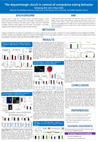
evoked IPSPs in a neuron negative for both D2R and ChR2 (right). (D)
Absence of dopamine D2 receptors increased Photostimulation mapping of local inhibitory input to a ChR2-negative
compulsive eating behavior in Light /dark box neuron. (E) Schematic for injection of ChR2-EYFP and implantation of a
test fiber-optic cannula in the CeA of D2R-Cre mice. (F) Total travel distance
in the open-field test during laser-on and laser-off periods for WT (n = 8)
and D2R-Cre (n = 6) mice. (G) Scheme for determining the effects of
optogenetic stimulation on performance in the 5-CSRTT. (H) Percentage
omission, accuracy and premature response during 15 min averaged over
the four test sessions of the 5-CSRTT performed with WT (n = 9) and
D2R-Cre (n = 6) mice. **P < 0.01 versus laser off, †P < 0.05 versus
corresponding WT value, genotype x light stimulation interaction in
premature response (%); F (1, 26) = 5.24, P = 0.0304 with two-way ANOVA
followed by Bonferroni test. (I) Time course omission (%), accuracy (%)
and premature response (%) in the 5-CSRTT as in (H). *P < 0.05 versus the
corresponding value for WT (unpaired Student’s t test). All data are
means ± SEM.
Fig 4. Analysis of compulsive eating behavior using optogenetics. (A)
Photostimulation of D2R-positive neurons in Schematic diagram of AAV-DIO-eNpHR3.0-EYFP injectionf into the CeA of
D2R-Cre mice and implanted fiber optic cannula into bed nucleus of the
the CeA regulates impulsive behavior stria terminalis (BNST). (B) Selective activation of D2R-expressing neurons
from CeA to BNST was achieved by 532nm laser during light/dark box
test for analysis of compulsive eating behavior. Test was done for 25min
divided into repeated laser off-laser on period (5min x 5 ). (C) Basal body
weight of WT and D2R-Cre mice for 7 days. (D) Basal food intake of WT
Fig 1. Compulsive-like eating behavior analysis with light/dark box test and D2R-Cre mice for 7 days. (E) Result of L/D Pre-test with WT and D2R-
in WT and Drd2 –/– mice. Cre mice. There was no difference in preference of each box between WT
(A) Experimental scheme of light/dark box test with WT and Drd2 –/– mice and D2R-Cre mice. (F) The number of crossover of L/D Pre-test with WT
to investigate phenotype of compulsive eating behavior. Mice were and D2R-Cre mice. There was no significant difference between WT and
induced strong preference for high-calorie palatable food for 14 days. (B) D2R-Cre mcie C-F; WT n=16, D2R-Cre n= 16. (G) Time spent in each box
Representative image of light/dark box test. (C) Basal body weight of WT during L/D test. (H) Food intake of normal chow in dark box or normal
and Drd2 –/– mice for 7 days. *** P < 0.001, student unpaired t-test. (D) chow/palatable food placed in light box for 25 min test session of WT
Basal food intake of WT and Drd2 –/– mice for 7 days. * P < 0.05, student and D2R-Cre mice. (I) Total food inatke in light box for 25 min. *** P
unpaired t-test. C-D; WT n=38, Drd2 –/– n=38. (E) Spent time in dark or < 0.001,# p <0.05, two-way ANOVA followed by bonferroni post-test. (J)
light box for 15min (test session) of WT and Drd2 –/– mice. ### P <0.0001; Food intake of WT and D2R-Cre in NC, PF groups during laser off of laser
WT versus Drd2 –/– in dark box, ††† P<0.0001; WT versus Drd2 –/– in light box, on period in L/D test (Palatable food: Two-way ANOVA followed by
*** P<0.0001; light box versus dark box in Drd2 –/– , interaction; F 1,158 =59.43, bonferroni test, genotype x light interaction: F 1,28 =7.32, P=0.0115; G-J; WT
P < 0.0001, Two-way ANOVA. (F) The number of crossover for 15min (test n=8, D2R-Cre n=8).
session) of WT and Drd2 –/– mice. Student t-test, ***p<0.0001. E-F; WT
n=39, Drd2 –/– n=42. (G) Spent time in light box in light/dark box pre-test CONCLUSION
and test. (H) Food intake of palatable food placed in the light box during
light/dark box test. Two-way ANOVA analysis, NC vs. PF ***P<0.0001, WT
vs Drd2 –/– . ###P < 0.0001, F 1,77 =15.55. (I) The number of crossover for We investigated novel dopaminergic circuitry regulating compulsive eating
15min (test session) of WT and Drd2 –/– . G-I; WT-NC n=23, WT-PF n=21,
KO-NC n=23, KO-PF n=23. behavior through dopamine D2 receptors (D2Rs). In the light/dark box test,
Fig. 3. D2R-expressing neurons in the CeA → BNST circuit regulate Drd2 –/– mice showed severe compulsive eating behavior in the aversive
Photostimulation of D2R-positive neurons in impulsive behavior. (A) Confocal images of patch-clamped neurons context. Central nucleus of the amygdala is known to be crucial for choice
(arrows; labeled red with Alexa Fluor 594) in the BNST of D2R-Cre mice
the CeA regulates impulsive behavior (n = 2) injected with AAV-DIO-ChR2-EYFP into their CeA. The neurons of incentive-reward and feeding behaviors. We previously identified that
were stained with antibodies to VGAT (blue). (B) Fraction of BNST D2R-expressing neurons in the CeA innervating to BNST regulate reward-
neurons examined (n = 45) that were positive or negative for related impulsivity. Selective optogenetic activation of D2R-expressing
photostimulation-induced IPSPs or VGAT immunoreactivity (n = 14). (C) neurons in CeABNST circuit induced deacrease of impulsivity (Kim et al.,
Representative light-evoked IPSCs recorded from VGAT(+) and VGAT(−)
neurons in the BNST. IPSC traces are the averages of five trials, measured PNAS, 2018). Here, we observed that optogenetic inhibition of
at a holding potential of −50 mV, and were low-pass-filtered (200 Hz). (D) CeABNST circuit enhanced compulsive eating behavior. We are
Amplitude of photostimulation-evoked IPSCs in VGAT(+) (n = 7) and currently investigating functional brain circuit regulating appetite control.
VGAT(−) (n = 3) BNST neurons (holding potential, −50 mV). Data were Our present findings may contribute to widen therapeutic strategies for
analyzed from two mice. (E) Schematic diagram of AAV-DIO-ChR2-EYFP
injection into the CeA and implantation of a fiber-optic cannula into the obesity and reward-related psychiatric disorders accompanied with eating
BNST of D2R-Cre mice (D2R-Cre CeA→BNST ) for analysis in F–H. (F) Total disorders.
travel distance per minute in the open-field test for WT (n = 5) and D2R-
Cre CeA→BNST (n = 3) mice during laser-off and laser-on (3 min, 5 Hz, 10
mW) periods. (G) Percentage omission, accuracy, and premature response REFERENCES
in the 5-CSRTT for WT (n = 17) and D2R-Cre CeA→BNST (n = 8) mice. *P
< 0.05 versus corresponding laser-off value; †P < 0.05 versus
corresponding WT; genotype × light stimulation interaction in premature 1. Bokyeong Kim et al, (2018) Proc Natl Acad Sci U S A. 115(45):E10730-E10739.
response (%): F (1, 30) = 7.96, P = 0.0084 with two-way ANOVA followed by
Bonferroni test. (H) Time course omission (%), accuracy (%), and 2. Oana Georgiana Rus et al, (2017) NeuroImage: Clinical 13, 246-255.
premature response (%) in the 5-CSRTT as in G. *P < 0.05 versus the 3. Paul M Johnson and Paul J Kenny, (2010) Nature Neuroscience 13, 635–641.
corresponding value for WT (unpaired Student’s t test). All data are
means ± SEM. 4. Akiyo Natsubori et al, (2017) Journal of Neuroscience 37 (10) 2723-273 .
5. Mike J.F. Robinson, Shelley M. Warlow and Kent C. Berridge. (2014) Journal
Inhibition of D2R-expressing neurons in of Neuroscience 34 (50) 16567-16580.
CeABNST circuit increased compulsive eating
ACKNOWLEDGEMENTS
behavior
Fig. 2. Photoactivation of D2R-positive neurons in the CeA regulates This work was supported by the Bio & Medical Technology Development
impulsive behavior. (A) Coronal section of the brain of a D2R-Cre mouse Program Grant no: 2016M3A9D5A01952412
injected into the CeA as indicated with ChR2-EYFP. (B)
Immunofluorescence staining for Cre as well as DAPI staining and EYFP
fluorescence (ChR2) for a section of the CeA of a D2R-Cre mouse injected Contact information
as in (A). Percent double-labeled cells out of the whole Cre-positive cell
population, and out of the whole ChR2-expressing cell population as First author: bobobo90@naver.com
counted from high-magnification confocal z-stacks (n = 7 slices per
animal from 2 mice). (C) Representative recordings of light- evoked action Corresponding author: jahyunb@korea.ac.kr
potentials in a ChR2-expressing D2R-positive neuron (left) and light-