
Page 77 - Q. Neuroscience
P. 77
TNF-α signaling and mitochondrial unfolded protein response
involved in the pathogenesis of hydrocephalus
Jiebo Zhu 1,2,3,*, Min Joung Lee 1,2,3,*, Hee Jin Chang 1,4, Yunseon Jang 1,2,3, Xianshu Ju 1,5, Jianchen Cui 1,5, Yu Lim Lee 1,5, Eunji Namgung 1,2,3, Dahyun Go 1,2,3,
Changjun Seo 1,2,3, Woosuk Chung 1,5,6, Eungseok Oh 1,4, Jun Young Heo 1,2,3,*
1 Department of Medical Science, 2 Department of Biochemistry, 3 Infection Control Convergence Research Center, 4 Department of Neurology, 5 Department of Anesthesiology and Pain Medicine, 6
Department of Anesthesiology and Pain Medicine Chungnam National University School of Medicine, Daejeon, South Korea, 35015
BACKGROUND Progressive ventriculomegaly induced by the abnormal flow of cerebrospinal fluid (CSF) lead to hydrocephalus, which accompanied by gliosis and
dysfunctional mitochondria. However, the underlying mechanism of neuroinflammation and mitochondrial dysfunction by ventriculomegaly were not fully understood.
AIM To identify the association between TNF-α signaling and UPRmt against ventricular expansion.
METHODS Generated hydrocephalic model by injected 25% kaolin into C57BL/6J mice cisterna magna with stereotactic surgery. Open field test and horizontal grid test to
detect the motor disturbances in kaolin induced hydrocephalus mouse model (KIHMM). Western blotting, ELISA, and immunofluorescence to assess the association
between TNF-α signaling and UPRmt in KIHMM.
RESULTS
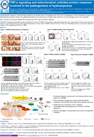
Figure 1 KIHMM showed ventricular enlargement Figure 2 KIHMM exhibited motor disturbances
A B C D E
A B C D E
F G (A, B) Movement activity was measured for 10 min in the
(A) Coronal sections showed ventricles in the open field test.
saline, 1-day, and 5-day group. Scale bar: (C) The neurological function was scored by a 5-point
200 μm. paradigm and plotted.
(B, C, D, E) The size of the lateral ventricle, (D, E, F, G) The hang time, successful steps, total steps,
dorsal 3rd ventricle, 3rd ventricle, and 4th and the ratio of successful to total steps, calculated for
ventricle. 30 s in the horizontal grid test.
Figure 3 TNF-α mediated neuroinflammation in KIHMM Figure 4 UPRmt activated in KIHMM Figure 5 Neuronal damaged in KIHMM
A B C D
A A
E F G
B C D B C
H I J
(A) The prefrontal cortex above the ventricles was stained for a marker of (A) The expression of Lonp1, Hsp60, and ClpP (A) The expression of PARP-1 and
microglia (Iba-1; red), and TNF-α (green). Scale bar: 20 μm. in the prefrontal cortex above the ventricles cleaved PARP-1 in prefrontal cortex
(B, C) The immunofluorescence intensity of Iba-1 and TNF-α was quantified. were analyzed by WB. above the ventricles were analyzed by
(D) The TNF-α level was analyzed by ELISA. (B, C, D) The intensity value of Lonp1, Hsp60, WB.
(E, F, G) The expression of Iba-1 and TNF-α by WB. and ClpP. (B, C) The intensity value of PARP-1
(H, I, J) The expression of p65 and P-p65 by WB. and cleaved PARP-1
CONCLUSION
1.Prominent dilatation ventricle and motor behavior defects were
observed in KIHMM.
2.TNF-α activated microglia and nuclear factor-κB (NF-κB)
signaling in the prefrontal cortex above the expanded ventricles.
3.Hsp60 induction preceded to TNF-α mediated
neuroinflammation accompanied by an increase of UPRmt
expression.
4.Neuronal damaged in the prefrontal cortex above the expanded
ventricles.
REFERENCES
1. Basati, S.; Desai, B.; Alaraj, A, et al. Cerebrospinal fluid volume measurements in hydrocephalic rats. J Neurosurg Pediatr 2012, 10 (4), 347-354.
2. Sosvorova, L.; Kanceva, R.; Vcelak, J, et al. The comparison of selected cerebrospinal fluid and serum cytokine levels in patients with multiple sclerosis and normal
pressure hydrocephalus. Neuro Endocrinol Lett 2015, 36 (6), 564-571.
3. Lackner, P.; Vahmjanin, A.; Hu, Q , et al. Chronic hydrocephalus after experimental subarachnoid hemorrhage. PLoS One 2013, 8 (7), e69571.
ACKNOWLEDGEMENTS The financial supports by the NRF, MSIP, and CNU. (2017R1A5A2015385, 2019M3E5D1A02068575, 2019R1F1A1059586)
Contact information Correspondence: junyoung3@gmail.com (J.Y.H.); Tel.: +82-042-580-8222