
Page 57 - Q. Neuroscience
P. 57
Development of levodopa induced dyskinesia mouse model
combining with mutant α-synuclein and MPTP
Eunji Namgung, 1,2,3,# Soo Jeong Kim, 1,3,# Min Jeong Ryu, 1 Yunseon Jang, 1,2,3 Min Joung Lee, 1,2,3 Xianshu Ju, 1,2 3 Jianchen Cui, 1,2,3 Yu Lim Lee, 1,2,3 Jiebo Zhu, 1,2,3 Dahyun Go,
1,2,3 Chang Jun Seo, 1,2,3 Woosuk Chung 4 and Jun Young Heo 1, 2, 3, *
1 Department of Biochemistry, 2 Department of Medical science, 3 Infection Control Convergence Research Center, Chungnam National University School of Medicine,
Daejeon 35015, Republic of Korea. 4 Department of Anesthesiology and Pain Medicine, Chungnam National University Hospital, # Co-first Author, * Coressponding Author
BACKGROUND For pre-clinical trial, the development of animal model which recapitulates Levodopa-Induced dyskinesia(LID) patient symptoms
should be preceded, at present the predominantly used model of LID is neurotoxin induced Parkinson’s disease(PD) animal model. MPTP and 6-
OHDA neurotoxic models display robust nigro-striatal degeneration but, construct validity are limited as the formation of LB inclusions is not a
common feature.
AIM To develop the recapitulate the LID patient symptoms, we generate the LID animal model combine with α-synuclein(α-syn) and MPTP.
METHODS Stereotactic surgery was performed with α-syn A53T virus in the SN area, and MPTP was administered by intraperitoneal injection.
Abnormal involuntary movement scale(AIMs) test was perfomed to identify the level of dyskinesia. TH positive neuron was confimed the DAB stain.
RESULTS
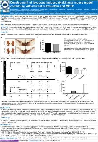
Figure 1. Levodopa-induced dyskinesia was not found in the animal model 1 week after sereotactic surgery with the mutated α-synuclein virus.
A B
(A) Immunohistochemical staining of the
10000 dopaminergic neurons in the STR and SN showed no
α-syn WT TH positive neuron 8000 clear differences between the two groups, a-syn
WT/A53T 1w group. (Scale bar: STR 200 μm and SN
100 μm)
6000
α-syn A53T 4000 0 a-syn WT 1w a-syn A53T 1w (B) Quantification of the dopaminergic neurons in the
2000
SN showed ~15% reduction between the two groups.
STR SN
Figure 2. The LID model was developed by injecting mitochondria complex 1 inhibition MPTP1 into mouse operated with α-synuclein A53T.
A B
α-syn A53T surgery L-dopa 2 treat / 1d , for a week Sacrifice 15000
α-syn A53T - . . . 0d 1d 4d 7d TH positive neuron 10000
21d
AIM test AIM test AIM test 5000
0
MPTP injection L-dopa 2 treat / 1d , for a week Control
MPTP . . . MPTP 20mg/kg α-syn A53T 3w
-7d 0d 1d 4d 7d C 200
AIM test AIM test AIM test AIM test total score 150
α-syn A53T surgery
α-syn + MPTP . . . MPTP injection L-dopa 2 treat / 1d , for a week Sacrifice 50
100
- -7d 0d 1d 4d 7d 0 PBS
21d α-syn A53T MPTP 20mg/kg A53T+MPTP
AIM test AIM test AIM test
(A) Develop combine mouse model timeline : performed stereotaxic surgery with α-syn A53T virus in the SN region, and administrated MPTP after two weeks later
and then treated LD twice a day for a week to induce LID. (B) Quantification of the dopaminergic neurons in the SN region between the three groups. (C) Results of
quantification of AIMs test on day 7
CONCLUSION
To develop mutant a-synuclein Levodopa-induced dyskinesia, which has not been established so far, we treated levodopa with a-synuclein A53T for
2 weeks after stereotactic surgery and identify movement with AIMs test, but only normal movement was confirm. Aggregated a-synuclein induces
mitochondria dysfunction by inhibiting mitochondria complex 1 and 4, and MPTP, which is a neurotoxin that inhibits mitochondria complex 1 and is
used as a PD and LID animal model so, the dyskinesia level in the combined model is more serious.
Futher study
By confirming the mitochondrial dysfunction of the dopamine neuron receptor, we identify that dyskinesia is caused by the effect of mitochondria on
the presynaptic pathway.
REFERENCES
1. Giráldez-Pérez et al. Models of α-synuclein aggregation in Parkinson’s disease. Acta Neuropathologica Communications 2014, 2:176
2.Lane E, Dunnett S. Animal models of Parkinson’s disease and L-dopa induced dyskinesia: how close are we to the clinic? Psychopharmacology
(Berl) 2008;199:303–312.A
3.K.F. Winklhofer, C. Haass. Mitochondrial dysfunction in Parkinson’s disease. Biochimica et Biophysica Acta 1802 (2010) 29–44
4.K.L. Norris et al. Convergence of Parkinm PINK1, and α-synuclein on Stress-induced Mitochondrial Morphological Remodeling. THE JOURNAL OF
BIOLOGICAL CHEMISTRY VOL. 290, NO. 22, pp. 13862–13874, May 29, 2015
ACKNOWLEDGEMENTS The financial supports by the NRF, MSIP, and CNU. (2017R1A5A2015385, 2019M3E5D1A02068575, 2019R1F1A105958612
Contact information Correspondence: junyoung3@gmail.com (J.Y.H.); Tel.: +82-042-580-8222