
Page 55 - Q. Neuroscience
P. 55
Oral administration of Proteus mirabilis, a gut bacterium linked to Parkinson’s disease,
alters the expression of a neurotrophin in the intestine of mice
Jun Heyok Kwak , Miran Jeong ,a,b,c , Jin Gyu Choi a,b,c , Eugene Huh , Jae-Won Lee , Im-Ho Lee , Dong-Hyun Kim a,b,c , Myung Sook Oh a,b,c , and
b
a,b
d
b
Jung-Hye Choi a,b,c,*
a Neurobiota Research Center (NRC), Kyung Hee University, Seoul, South Korea, Department of Life and Nanopharmaceutical Sciences, Kyung Hee University, Seoul, South Korea , c
b
College of Pharmacy, Kyung Hee University, Seoul, South Korea, Medical Science of Meridian, Graduate School, Kyung Hee University, Seoul, South Korea
d
Abstract Results
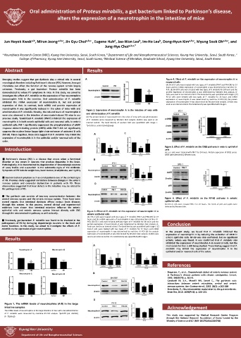
Emerging studies suggest that gut dysbiosis play a critical role in several Figure 4. Effect of P. mirabilis on the expression of neurotrophin A in
neurological disorders including Parkinson’s disease (PD). However, how gut neuronal cells
microbiota can affect the function of the nervous system remain largely (A) SH-SY5Y cells were treated with two types of P. mirabilis (PM-T and PM-CM) for 24
hours and the mRNA expression of neurotrophin A was determined by real-time RT-
unknown. Previously, a gut bacterium Proteus mirabilis has been PCR. (B) SH-SY5Y cells were treated with two types of P. mirabilis for 24 hours and the
demonstrated to induce PD symptoms in mice. In this study, we aimed to protein expression of neurotrophin A was determined by Western blot analysis. β-
investigate the effect of P. mirabilis on the expression of four neurotrophins Actin was used as an internal control. The band density was quantified with Image J. (C)
PC-12 cells were treated with two types of P. mirabilis for 24 hours and mRNA
(neurotrophin A-D) in the intestine. Oral administration of P. mirabilis expression of neurotrophin A was determined by real-time RT-PCR (D) the protein
inhibited the mRNA expression of neurotrophin D, but not protein expression of neurotrophin A was determined by Western blot analysis. β-Actin was
expression of that. In contrast, both mRNA and protein expression of used as an internal control. The band density was quantifiedwith Image J.
neurotrophin A was significantly reduced in the colon of mice with oral Figure 2. Expression of neurotrophin A in the intestine of mice with
administration of P. mirabilis. Notably, the reduced levels of neurotrophin A oral administration of P. mirabilis
were also observed in the intestine of neurotoxin-induced PD mice in our (A) The protein levels of neurotrophin A in the colon of mice with oral administration
previous study. Tyndallized P. mirabilis (PM-T) inhibited the expression of of P. mirabilis were measured by Western blot analysis. β-Actin was used as an
neurotrophin A in both enteric epithelial cells and neuronal cells. In enteric internal control. The band density of western blot was quantified with Image J.
epithelial cells, PM-T significantly suppressed the phosphorylation of cAMP *p<0.05 (vs. control)(n=5/group).
response element-binding protein (CREB) while PM-T did not significantly
suppress the nuclear factor kappa-light-chain-enhancer of activated B cells
(NF-kB). Taken together, these data suggest that P. mirabilis may inhibit the
expression of neurotrophin A in the epithelial and/or neuronal cells of the
colon.
Introduction Figure 5. Effect of P. mirabilis on the CREB pathway in enteric epithelial
cells
(A) STC-1 cells were treated with PM-T for 24 hours. Protein expression of CREB and p-
CREB were detected by Western blot.
■ Parkinson’s disease (PD) is a disease that occurs when a functional
disorder or loss occurs in neurons that produce dopamine in the brain.
Pathologically, it is characterized by degeneration of dopaminergic neurons
of Lewy bodies and a-synuclein in the substantia nigra of the midbrain.
Symptoms of PD include weight loss, hand tremor, bradykinesia, and rigidity.
■ Gastrointestinal symptom such as constipation is one of the earliest signs
of PD. Previous study suggested correlation between changes in the enteric
nervous system and colorectal dysfunction in patients with PD. These
observations suggested that local defects in the intestine may be related to
the pathogenesis of PD.
■ The gut-brain axis consists of two-way communication between the
central nervous system and the enteric nervous system. There have been Figure 6. Effect of P. mirabilis on the NF-kB pathway in enteric
several reports that intestinal dysbiosis affects various brain diseases epithelial cells
through this system. In this process, several clinical and experimental (A) STC-1 cells were treated PM-T for 24 hours. The levels of p65 and p-p65 were
evidences have shown that intestinal microbes influence the enteric detected by Western blot.
epithelial cells and enteric nervous system interact directly with CNS Figure 3. Effect of P. mirabilis on the expression of neurotrophin A in
through the neuroendocrine pathway as well as locally. enteric epithelial cells
(A) STC-1 cells were treated with two types of P. mirabilis (PM-T and PM-CM) for 24
■ Previously, gut bacterium P. mirabilis was found to be involved in the hours and the mRNA expression of neurotrophin A was determined by real-time RT-
PCR. (B) STC-1 cells were treated with two types of P. mirabilis for 24 hours and the
pathogenesis of PD by damaging dopaminergic neurons in the brain and protein expression of neurotrophin A was determined by Western blot analysis. β- Conclusion
motor functions. In this study, we aimed to investigate the effects of P. Actin was used as an internal control. The band density was quantified with Image J. (C)
mirabilis on the expression of gut neurotrophins. Caco-2 cells were treated with two types of P. mirabilis for 24 hours and mRNA
expression of neurotrophin A was determined by real-time RT-PCR (D) the protein In the present study, we found that P. mirabilis inhibited the
expression of neurotrophin A was determined by Western blot analysis. β-Actin was expression of neurotrophin A by reducing the activation of CREB in
Results used as an internal control. The band density was quantifiedwith Image J. enteric epithelial cells. NF-kB was also confirmed, but no significant
activity reduce was found. It was confirmed that P. mirabilis also
inhibited the expression of neurotrophin A in neuronal cells, but the
mechanism for that is still being studied. These finding suggest that P.
mirabilis may inhibit the expression of neurotrophin A in the
Neurotrophin A 1.5 Neurotrophin B epithelial and/or neuronal cells of the colon.
1.5
Relative mRNA expression 1.0 0.5 * Relative mRNA expression 1.0 0.5 References
0.0
Control PM 0.0 Control PM - Singaram, C., et al., Dopaminergic defect of enteric nervous system
Neurotrophin C 1.5 Neurotrophin D in Parkinson’s disease patients with chronic constipation. Lancet,
1.5
1995. 346(8979): p. 861-4.
Relative mRNA expression 1.0 0.5 Relative mRNA expression 1.0 0.5 * - Carabotti M, S.A., Maselli MA, Severi C., The gut-brain axis:
interactions between enteric microbiota, central and enteric
nervous systems. Ann Gastroenterol., 2015. 28(2): p.203-209
- Strandwitz, P., Neurotransmitter modulation by the gut microbiota.
0.0
Control PM 0.0 Control PM Brain Res, 2018. 1693(Pt B): p. 128-133.
Figure 1. The mRNA levels of neurotrophins (A-D) in the large
intestine samples. Acknowledgement
The mRNA levels of neurotrophins in the large intestine of mice with oral administration
of P. mirabilis were measured by real-time RT-PCR analysis. *p<0.05 (vs. control)
(n=5/group). This study was supported by Medical Research Center Program
through the National Research Foundation of Korea funded by the
Ministry of Science and ICT (NRF-2017R1A5A2014768)
Kyung Hee University
Department of Life and Nanopharmaceutical Sciences