
Page 51 - Q. Neuroscience
P. 51
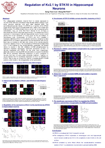
Regulation of Kv2.1 by STK10 in Hippocampal
Neurons
Sang Yeon Lee , Kang-Sik Park 1,2
1
1 Department of Biomedical Science, Graduate school, and 2 Department of Physiology, College of Medicine, Kyung Hee University, Seoul, South Korea
,
Abstract 4. Knockdown of STK10 inhibits somato-dendritic clustering of Kv2.1
The voltage-gated potassium channel Kv2.1 is widely expressed in
neurons of the CNS and is localized in high density surface clusters on the
soma, proximal dendrites, and axon initial segment (AIS). The
serine/threonine-protein kinase 10 (STK10) is a member of the STE20
kinase family and is able to phosphorylate ezrin, radixin, and moesin (the
ERM proteins). The ERM proteins have a critical role in reorganizing the
cytoskeleton. Here, we show that Kv2.1 channel in the rat brain is
associated with STK10 using mass spectrometry. The endogenous STK10
expression in heterologous cells and hippocampal neuron reveals overall
puncta form in the cytoplasm or column form on the cell surface. In
heterologous cells, co-expression of STK10 and Kv2.1 resulted in
increased trafficking of Kv2.1 to the plasma membrane compared to trials
without STK10. In cultured hippocampal neurons transfected with STK10,
there was an increase in the size and number of the somato-dendritic Immunoblot demonstrating the efficacy of STK10 shRNA (A). The blot was probed with anti-V5 antibody. Hippocampal neurons
membrane clusters of Kv2.1. The knockdown of STK10 demonstrated that were transfected with shRNA-STK10-RFP, Kv2.1-GFP, shRNA-NC-RFP in DIV 14-17. Knockdown of STK10 using shRNA-
Kv2.1 is not localized in the somato-dendritic membrane, but instead STK10-RFP blocked the somato-dendritic cluster of Kv2.1 (B) compared to endogenous Kv2.1 non-transfected with shRNA-
STK10-RFP (C). Likewise, shRNA-STK10-RFP decreased clusters of Kv2.1-GFP (D) compared to Kv2.1-GFP co-expressed with
retains its clusters in the AIS. Inhibition of ERM phosphorylation by shRNA-NC-RFP (E).
rapamycin treatment suppressed membrane expression of Kv2.1 in 5. Rapamycin inhibits cytoskeleton reorganization by suppressing ERM
neurons transfected with STK10. On the other hand, interestingly, phosphorylation
glutamate treatment inducing ERM phosphorylation in neurons transfected
with STK10 increased membrane expression of Kv2.1 compared to
neurons that have not gone under glutamate treatment. Together our
results indicate that the membrane expression of Kv2.1 is regulated by
STK10. This is done so through the method of phosphorylation of ERM
proteins, which results in the reorganization of the cytoskeleton.
1. Localization of endogenous STK10 in two distinct types
We treated rapamycin on hippocampal neurons to confirm the cluster change of Kv2.1 when ERM phosphorylation by STK10 was
suppressed. Hippocampal neuron treated with DMSO (A), rapamycin (10uM) (B) for 15min. Hippocampal neuron transfected with
STK10-V5 treated with DMSO (C), rapamycin (D). STK10-V5 treated with rapamycin decreased cluster size (E) and number (F) of
soma of endogenous Kv2.1 in neurons.
6. Glutamate receptor-mediated ERM phosphorylation regulates
Kv2.1 expression
STK10 is uniquely column-shaped on the membrane (A, C, E) whereas its expression is uniformly expressed in the
cytoplasm (B, D, F) in HEK293 cells (A, B), COS-7 cells (C, D) and hippocampal neurons (E, F).
2. Subcellular localization of Kv2.1 and STK10 in heterologous
cells
We treated glutamate, which is known to induce ERM phosphorylation. Hippocampal neuron treated with D.W. (A), glutamate
(10uM) (B) for 30min. Hippocampal neuron transfected with STK10-V5 treated with D.W. (C), glutamate (D). STK10-V5 treated
with glutamate increased cluster size (E) and number (F) of soma of endogenous Kv2.1 in neurons compared to non-transfected
Location and expression of Kv2.1-RBG4, STK10-V5 in HEK293 cells (A, B, C) and COS-7 cells (D, E, F). STK10 may neurons treated with glutamate.
regulate the plasma membrane expression of Kv2.1. The Kv2.1 channel proteins co-expressed with STK10 showed an
increase of biotinylated fraction compared to that the Kv2.1 alone (G). Kv2.1 co-expressed with STK10 resulted in an
increased biotinylated fraction level of 28% compared to biotinylated fraction levels of Kv2.1 alone (H). 7. The membrane expression of Kv2.1 is regulated by STK10,
which is cytoskeleton reorganization by ERM phosphorylation
3. Regulation of the expression of Kv2.1 to the membrane by STK10
in hippocampal neurons
Hippocampal neurons were transfected with STK10-V5, Ezrin-WT-Flag, Ezrin-T567D-Flag, Ezrin-T567A-Flag in DIV 17. The
neuron transfected with STK10-V5 (B) increased phosphor ezrin compared to non-transfected neuron (A). Ezrin-WT-Flag induced
the large somato-dendritic clusters of Kv2.1 in plasma membrane (C). Ezrin-T567D-Flag (D), conformational change mutant, and
Ezrin-T567A-Flag (E), Ezrin dephosphorylation mutant, decreased clusters of Kv2.1. Theses statistics of Kv2.1 clusters size (F)
and number (G) revealed in graphs.
Conclusion
STK10 is complexed with Kv2.1 channel in rat brain.
The endogenous STK10 expression in heterologous cells and hippocampal
neuron reveals overall puncta form in the cytoplasm or column form on the cell
surface.
Hippocampal neurons were transfected with Kv2.1-GFP, STK10-V5 in DIV 14-17. Kv2.1 localizes high density clusters in the
soma, proximal dendrite and AIS (A, H). STK10-V5 increases cluster size and number of soma (C, D, J, K) , AIS (E, F, G, L) of
endogenous Kv2.1 (B) and Kv2.1-GFP (I) in neurons. STK10 activated by active RhoA affects the somato-dendritic membrane
clusters of Kv2.1 by cytoskeleton reconstruction by phosphorylating ERM.