
Page 37 - Q. Neuroscience
P. 37
MicroRNA-24-3p regulates neuronal differentiation by modulating
hippocalcin expression
Min-Jeong Kang, Shin-Young Park*, and Joong-Soo Han*
1 Department of Biomedical Sciences, Graduate School of Biomedical Science & Engineering, and Biomedical
2
Research Institute and Department of Biochemistry & Molecular Biology, College of Medicine, Hanyang University,
222 Wangsimni-ro, Seongdong-gu, Seoul 04763, Republic of Korea.
E-mail : jshan@hanyang.ac.kr, ttokttok@hanyang.ac.kr, TEL : 82-2-2220-0623, 82-2-2220-0610.
ABSTRACT INTRODUCTION
Hippocalcin (HPCA) is a neuron-specific calcium-binding protein predominantly expressed in the MicroRNAs are small, highly conserved non-coding RNA molecules of approximately 22 nucleotides.
nervous system. In the present study, we demonstrate that HPCA regulates neuronal differentiation. They can modulate gene expression through complementary base pairing of the seed sequence located
We observed that the expression level of HPCA was increased during neuronal differentiation. in the 3’UTR of the target mRNA, leading to the translational suppression and/or destabilization of the
Depletion of HPCA inhibited both neurite outgrowth and synaptophysin (SYP) expression. shRNA- target mRNAs [1]. It is widely accepted that a single miRNA has the potential to inhibit the expression of
mediated knockdown of HPCA in the hippocampal dentate gyrus exhibited manic-like behavior, hundreds of target mRNAs and, conversely, individual mRNAs are commonly targeted by multiple
including hyperactivity, decreased anxiety-like behavior, reduced depressive-related behavior, and miRNAs [2]. Hence, miRNAs serve as key regulators in various biological processes, such as
impaired learning and memory. Furthermore, HPCA depletion reduced the levels of synaptic plasticity- proliferation, differentiation, apoptosis, metabolism, and development [3].
related proteins. Thus, HPCA regulates neuronal differentiation both in vitro and in vivo. Interestingly, Hpca is a high-affinity calcium-binding protein expressed most
we also found that the expression of HPCA was modulated by miR-24-3p. We showed that co- abundantly in pyramidal cells of the hippocampal CA1 region [4].
transfection of a plasmid containing the miR-24-3p binding site from the 3'-untranslated region of the It belongs to the family of EF-hand-containing neuronal calcium
HPCA gene and an miR-24-3p mimic effectively reduced luminescence activity. miR-24-3p expression sensor proteins that possess a Ca2+/myristoyl switch allowing
was decreased during differentiation, suggesting that the decreased expression level of miR-24-3p translocation to membranes in response to increased cytosolic
might have upregulated mRNA expression of HPCA. As expected, upregulation of miR-24-3p by an Ca2+ concentration [5]. Our previous study showed that HPCA
miRNA mimic led to reduced HPCA expression, accompanied by diminished neuronal differentiation. In increases NeuroD expression, resulting in neurite outgrowth
contrast, downregulation of miR-24-3p by an antisense inhibitor promoted neurite outgrowth as well as during the differentiation of H19-7 cells [6]. Moreover, it was found
levels of SYP expression. Taken together, these results suggest that miR-24-3p is an important miRNA to promote neuronal differentiation through activation of the
that regulates neuronal differentiation by controlling HPCA expression.
PKCα/PLD1/SHP1 cascade, leading to the inhibition of astrocytic
Keywords: Hippocalcin, miR-24-3p, neuronal differentiation, Synaptophysin, SH-SY5Y cells differentiation in cortical neural stem cells [7].
RESULTS
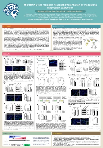
Fig. 1 Differentiation induced expression of HPCA in SH-SY5Y cells Fig. 3 HPCA deficiency in DG exhibited manic-like behavior and Fig.5 Identification of miR-24-3p as a potential upstream regulator of
inhibited hippocampal neurogenesis. HPCA
(A, B) HeLa cells were
transiently transfected with a
control miRNA mimic or an
miR-24-3p mimic for 2 days.
(C, D) Cells were transfected
with a control miRNA inhibitor
or an miR-24-3p inhibitor for 3
days. The graph shows mean
densities as fold increases
from three independent
experiments (mean ± SEM).
**P < 0.01, ***P < 0.001
compared with the control.
(A, B) SH-SY5Y cells were induced to differentiate by adding 50 μM RA for the
indicated number of days. (A) The mRNA levels of presynaptic marker, SYP and HPCA
were determined by RT-qPCR. (B) Proteins were analyzed by western blotting with Fig.6 miR-24-3p regulates neuronal differentiation, presumably
anti-HPCA, anti-SYP, and anti-calnexin antibodies. (C) Cells were stained with DAPI by reducing HPCA expression in SH-SY5Y cells
(blue) and an anti-SYP antibody (green) to visualize neurite extensions. Scale bar, 100
μm. (D) Neurite lengths were measured in randomly selected areas in three
independent experiments. **P < 0.01, ***P < 0.001 compared with day 0 of
differentiation (means ± SEM).
Fig. 2 Effect of HPCA on the neuronal differentiation of SH-SY5Y
cells
(A) Design of the lentiviral construct expressing mouse Hpca shRNAs. (B) Timeline of
experimental paradigm. (C) Fixed brains 1 month after infusion of the virus were
immunostained with an anti-GFP antibody (green) and DAPI (blue) to confirm the
localization of lentivirus infection in the DG. Scale bar, 80 μm. (D) Proteins were analyzed
by western blotting with anti-calnexin, anti-PSD95, anti-NMDAR2C, anti-SHANK1, and
anti-calnexin antibodies. (E, F) Locomotor activity. (G) Contextual fear memory. (H)
NSFT. (I) FST. Data are mean ± SEM.
Fig. 4 Validation of HPCA as a direct target of miR-24-3p
(A) Mature miR-24-3p levels during the differentiation of SH-SY5Y cells were quantified
using TaqMan RT-qPCR. (B, C) Differentiation was induced in SH-SY5Y cells transfected
with a control miRNA mimic or an miR-24-3p mimic. (D) Fixed cells were stained with an
anti-SYP antibody (green) and DAPI (blue). Scale bar, 100 μm. (E) Neurite lengths were
measured in randomly selected areas from three experiments (mean ± SEM). *P < 0.05,
**P < 0.01, ***P < 0.001 compared with the control.
Fig.7 Inhibition of miR-24-3p promoted the neuronal differentiation of
SH-SY5Y cells
(A, B) SH-SY5Y cells were transiently transfected with control siRNA or HPCA siRNA,
and then incubated for 7 days after the addition of RA. (C) Fixed cells were stained with
an anti-SYP antibody (green) and DAPI (blue). Scale bar, 100 μm. (D) Neurite lengths (A) Map of representative bicistronic firefly/Renilla luciferase (FLuc/RLuc) plasmids
were measured in randomly selected areas from five independent cultures. (E, F) SH- containing HPCA mRNA 3'UTRs with putative miR-24-3p binding sites (WT) or mutated
SY5Y cells were transduced with pMSCV-IRES-EGFP or pMSCV-HPCA-Myc-IRES- binding sites (Mut). (B) A control miRNA mimic or an miR-24-3p mimic was co-
EGFP and induced to differentiate for 7 days. (G) Immunofluorescence was used to transfected with HPCA-3'UTR-WT or HPCA-3'UTR-Mut into HeLa cells. (C) A control (A, B) SH-SY5Y cells were transfected with a control miRNA inhibitor or an miR-24-3p
visualize SYP (green) at day 7 of differentiation, with or without overexpression of miRNA inhibitor or an miR-24-3p inhibitor was co-transfected with HPCA-3'UTR-WT or inhibitor and were allowed to differentiate for 5 days. (C) Immunofluorescence was used
HPCA. Nuclei were stained with DAPI (blue). (H) Neurite lengths were measured in HPCA-3'UTR-Mut into HeLa cells. The relative activity of luciferase in control miRNA to visualize SYP (green) at day 7 of differentiation. Scale bar, 100 μm. (D) Neurite lengths
randomly selected areas from five slides of each condition. **P < 0.01, ***P < 0.001 inhibitor-transfected cells was set to 1.0. **, P < 0.01; ***, P < 0.001; n.s., not significant. were measured in random areas from five cultures (mean ± SEM). **P < 0.01, ***P
compared with the control (mean ± SEM). < 0.001 compared with the control miRNA inhibitor.
CONCLUSION REFERENCES
1. Djuranovic S, Nahvi A, Green R (2012) miRNA-mediated gene silencing by translational repression followed by mRNA deadenylation and decay.
miR-24-3p as a putative regulator of Science 336 (6078):237-240.
2. Esquela-Kerscher A, Slack FJ (2006) Oncomirs - microRNAs with a role in cancer. Nat Rev Cancer 6 (4):259-269.
HPCA during neuronal differentiation. 3. Luo W,Nie Q, Zhang X (2013) MicroRNAs involved in skeletal muscle differentiation. J Genet Genomics 40 (3):107-116.
4. Kobayashi, M., Takamatsu, K., Saitoh, S., and Noguchi, T. (1993). Myristoylation of hippocalcin is linked to its calcium-dependent membrane
miR-24-3p is an important miRNA association properties. J Biol Chem 268(25), 18898-18904.
5. 21. O'Callaghan DW, Tepikin AV, Burgoyne RD (2003) Dynamics and calcium sensitivity of the Ca2+/myristoyl switch protein hippocalcin in living
that regulates neurite outgrowth and cells. J Cell Biol 163 (4):715-721.
neuronal differentiation through 6. Oh, D.Y., Cho, J.H., Park, S.Y., Kim, Y.S., Yoon, Y.J., Yoon, S.H., et al. (2008). A novel role of hippocalcin in bFGF-induced neurite outgrowth of
controlling HPCA expression. H19-7 cells. J Neurosci Res 86(7), 1557-1565.
7. Park, S. Y., Yoon, S. N., Kang, M. J., Lee, Y., Jung, S. J., & Han, J. S. (2017). Hippocalcin promotes neuronal differentiation and inhibits astrocytic
differentiation in neural stem cells. Stem cell reports, 8(1), 95-111.