
Page 39 - Q. Neuroscience
P. 39
XPn interacts with microtubules to impair the
function of tau for neurite degeneration
Hyejin Park*, Youngwon Kim*, Yong-Keun Jung
School of Biological Science, Seoul National University, Seoul 08826, Korea
BACKGROUND AIM
X proteins (XPs) play a key role in nucleotide metabolism via nucleotide phosphoryl In this study, we aim to identify the function of XPn.
exchange. A new member of this family, XPn, has recently been identified, but its
function remains largely unknown.
METHODS
Cell culture, cell death assessment and DNA transfection
HEK293T (human embryonic kidney) and SH-SY5Y (human neuroblastoma) cells were cultured in DMEM (Hyclone) supplemented with 10% (v/v) fetal bovine serum (Hyclone). To differentiate SH-SY5Y, cells were treated with 10 mM retinoic acid (RA) in the medium containing 1 % serum.
Transfection in HEK293T and SH-SY5Y cells were performed using the Polyfect reagent (Qiagen) and cell death in SH-SY5Y was assessed by counting the number of GFP and EtHD-positive cells after staining with 0.5 mM ethidium homodimer (EtHD; Molecular probes) for 15 min. Primary cortical
or hippocampal neurons from mouse embryos (E16 or E18) were prepared and seeded on poly- L -lysine (0.01% in 100 mM borate buffer pH 8.5)-coated glass cover slips. The cells were maintained in neurobasal medium containing 2% B-27 supplement (Invitrogen) and 0.5 mM L -glutamine. Half of the
medium was exchanged every 3 days. Primary neurons were transfected using the LipofectAMINE TM 2000 reagent (Invitrogen) according to the manufacturer’s instructions.
Western blotting and antibodies
Cells were lyzed in ice-cold lysis buffer (50 mM Tris-Cl, pH 7.4, 150 mM NaCl, 1% Triton X-100, 1 mM sodium orthovanadate, and a mixture of protease inhibitors) and sonicated briefly. Cell lysates were clarified by centrifugation, separated by SDS-PAGE and blotted onto PVDF membrane. The
blots were blocked for 1 h with 5% BSA and incubated with following antibodies: a-tubulin, b-tubulin, b-actin (Sigma) and GFP (Santa Cruz Biotechnology). Polyclonal rat anti-XPn antibody was generated by immunizing rats with purified His-XPn ΔP1 protein, following standard immunization
procedures. Membranes were washed and incubated for 1 h with peroxidase-conjugated anti-mouse, rabbit or rat antibody and visualized using ECL detection system.
Production of recombinant protein and pull-down assay
Pull down assay was previously described. cDNA fragment encoding the full length human XPa, XPb, XPc, XPn and deletion mutants (ΔP1, ΔP2, ΔP1P2) were inserted into pET-28a vector. All fragments were confirmed by DNA sequencing. E.coli BL21 cells transformed with each plasmid were
cultured to reach an OD600 of 0.6, before induction with 0.5 mM IPTG. After incubation for 16 h at 16 °C, cells were harvested and lysed by sonication. His-fused XPns or XPn deletion mutants were purified form the cell lysates by adsorption onto Ni-NTA chelating agarose CL-6B (Peptron). To find
out the interaction proteins of XPn, 1 mg of mouse brain lysates were incubated with XPa, XPb, XPc or XPnΔP1 deletion mutant protein bounded to Ni-NTA chelating agarose overnight at 4 °C. After 5 intensive washing, proteins were eluted, separated on SDS-PAGE and stained with Coomassie
blue. The three protein bands of interest were excised from the gel and analyzed by mass spectrometry. To demonstrate the interaction between XPn and tubulin, His-XPn or deletion mutants conjugated to Ni-NTA agarose were incubated with mouse brain extract in TBS buffer (25 mM Tris-Cl, pH7.4,
150mM NaCl) with 1 mM DTT, 1 mM PMSF and 1 % Triton X-100 for 1 h at 4 °C. The beads were pulled-down by centrifugation and washed, and then subjected to western blotting.
Immunocytochemistry
Cells were fixed 4% paraformaldehyde (PFA) (Sigma) for 15 min, rinsed with PBS and permeablized with 0.1% Triton X-100 for 5 min. After blocking with PBS containing 5% BSA, cells were double-stained with XPn (1:100) and α-tubulin (1:500) for 2 h and washed with PBS before the incubation
with FITC-conjugated or TRITC-conjugated secondary antibodies (Jackson Laboratory, Inc.). After rinsing with PBS, the cover slips were placed with a mounting solution (Sigma) and samples were observed on a confocal laser scanning microscope (LSM510, Carl Zeiss, Inc.).
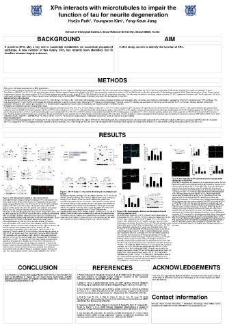
RESULTS
XPn FL
XPn
XPn
XPn
XPn ΔP2 XPn ΔP1P2 XPn
70 XPn FL XPn
70 XPn ΔP2
XPn 42 XPn ΔP1P2
XPn ΔP1
70 XPn ΔP1
FL
XPn
XPn FL siXPn siXPn/Aβ
XPn
XPn
siXPn siXPn
XPa XPn ΔP2 XPn ΔP1P2
XPn XPn ΔP2 XPn ΔP1P2 XPn ΔP1 XKn ΔP2 XKn ΔP1P2
Figure 4.Down-regulation of XPn prevents amyloid-β-induced neurite
degeneration and cell death
(A and B)XPn expression is increased by Aβ in hippocampal neurons. Primary
XPn ΔP1
XKn ΔP1 hippocampal neurons were prepared and then left untreated or exposed to the
XPn XPn increased dose of Aβ 42 at day 14 in vitro (DIV). After 24 h, cell extracts were
FL subjected to western blot analysis using XPn antibody (A). Densitometric
analysis of XPn levels on the blots shown in (A). Bars represent mean values
XPn
Figure 2. XPn P2 domain is required for the binding to microtubules and ±SD (n=3). *P<0.05, ** P< 0.005 (B). (C)Enhanced immunoreactivityagainst
tubulin XPn in the hippocampal neurons exposed to Aβ 42 . Mouse hippocampal
(A)Diagram depicting full-length XPn and deletion mutants (ㅿ). P1 and P2 are neurons were incubated with 5 µM Aβ 42 for 24 h. Expression of XPn was
Figure 1. XPn interacts with tubulin in vitro and in neurons domains showing homology with X kinases. (B)Pull-down assay showing the XPn examined with immunostaining using XPn antibody and hoechst dye.
(A and B) Pull-down assays showing the binding of XPn to a/b-tubulin in the binding of XPn deletion mutants to tubulin. Mouse brain lysates were (D)Reduced expression of XPn prevents Aβ 42 -mediated neurite degeneration.
mouse brain extract. Mouse brain extracts were subjected to the pull-down incubated with purified His-XPn or deletion mutant proteins (10 mg) coupled Mouse hippocampal neurons were transfected with GFP and scrambled (Scr)
assays using purified His-XPa, His-XPb, His-XPc or His-XPn ΔP1 protein(20 onto Ni-NTA agarose and the proteins bound to the beads were detected by or siXPn RNA for 48 h and then were left untreated or incubated with 5 µM
Aβ 42 . After 24 h, cells were then fixed with 4% paraformaldehyde (PFA) and
mg)(A) or His-GFP or His-XPn protein (10 mg) (B) coupled onto Ni-NTA western blotting using a-tubulin and His antibodies. The arrows on the right
agarose and the proteins bound to the agarose were analyzed with western indicate the apparent molecular mass of each His-tagged XPn mutant and FL examined under a confocal microscope. Scale bars, 20 mm. (E) Neurons
showing degeneration including the beaded and dystrophic neurites were
blotting using the indicated antibodies.(C) Overlay assays showing the asterisks indicate the nonspecific band of XPn.(C) Microtubules counted. Data are represented as the mean ±SD from three independent
interaction of XPn with a/b-tubulin in the brain extract. Purified XPn protein (10 cosedimentation assay of XPn deletion mutants. Purified His-XPn or His-XPn Figure 3.XPn overexpression induces neurite degeneration and
mg) was separated by SDS-PAGE and transferred to nitrocellulose membrane. deletion mutant proteins were incubated with or without the prepolymerized promotes neuronal death experiments and P values were calculated using t-test. *P<0.05. (F)Reduced
expression of XPn prevents Aβ 42 -inducedneuronal death. Mouse hippocampal
After an overnight incubation with mouse brain extract, the membrane was microtubules and the reactions were subjected to centrifugation to separate (A and B)Ectopic expression of XPn increases neurite degeneration in cell line, HT22were transfected with the GFP and scrambled (Scr) or siXPn
probed by western blotting using a/b-tubulin, b-actin and His-antibodies. (D and supernant (S) and pellet (P). Equal amounts of the supernant and the pellet differentiated SH-SY5Y with RA. SH-SY5Y cells were treated with 10 mM RNA for 24 hand then were left untreated or incubated with 5 µM Aβ 42 for 24 h.
E) Endogenous XPn binds to a/b-tubulin. Mouse brain extracts were were analyzed by western blotting using His and α-tubulin antibodies. RA for 7 days, transfected with the GFP-tagged XPn or XPn deletion After staining with EtHD, cell death was quantified by counting the number of
immunoprecipitated (IP) with XPn (D) or a-tubulin antibody (E) and the mutants. After additional 24 h, GFP-positive cells were observed under both GFP and EtHD-positive cells amongtotal GFP-positive cells. Bars
immunoprecipitates were analyzed by immunoblotting using the indicated fluorescence microscope. Scale bars, 20 mm (A). The cells showing neurite represent mean values ±SD (n = 3).P values were calculated using t-test. **
antibodies. (F) XPn associates with microtubules in vitro. Purified His-GFP and degeneration were counted. Data are represented as the mean ±SD from P< 0.005.
His-XPn proteins were incubated with (+MT) or without (-MT) the three independent experiments. P values were calculated using t-test.
prepolymerized microtubules. After centrifugation, equal amounts of the *P<0.05, ** P< 0.005. (C and D) XPn induces degeneration of neurites in
supernant (S) and the pellet (P) of the incubation mixtures were processed by cultured hippocampal neurons. 14-day-in-cultured hippocampal neurons
SDS-PAGE and proteins were then stained with CoomassieBrillant Blue.(G) were transfected with GFP, full-length GFP-XPn and deletion mutants of
XPn associates with microtubules in cells. HEK293T cells were transfected XPn. Neurons were then fixed with 4% paraformaldehyde (PFA) and
with GFP or XPn for 24 h and cell lysates were incubated with 50 mM taxol in examined under a confocal microscope. Scale bars, 20 mm (C). The cells
the presence of GTP. The samples were then spun through a cushion buffer showing numerous signs of degeneration, including the beaded and
containing 40% glycerol at 100,000 g for 45 min. After sedimentation, the dystrophic neurites were counted. Data are represented as the mean ±SD
precipitates (Pellet) and supernatants (Sup) were subjected to western blot from five independent experiments and P values were calculated using t-test.
analysis using the indicated antibodies. (H) Co-localizion of XPn with tubulin in *P<0.05, ** P< 0.005 (D). (E)XPn P2 domain is essential for XPn-mediated
cortical neurons. Primary cortical neurons were prepared from embryonic cell death. SH-SY5Y cells were transfected with the GFP-XPn or XPn
mouse (E-16) and cultured for 10 days in neurobasal medium. The neurons mutants for 36 h. After staining with EtHD, cell death was quantified by
were then immunostained with XPn and a-tubulin antibodies. Bar, 20 mm. counting the number of both GFP and EtHD-positive cells among total GFP-
positive cells. Bars represent mean values ±SD (n = 3).P values were
calculated using t-test. *P<0.05, ** P< 0.005 (left). The expression level of
GFP-fusion proteins was examined by western blotting using the GFP
antibody. The arrows on the right indicate the apparent molecular mass of
GFP-tagged XPn and deletion mutants (right).
CONCLUSION REFERENCES ACKNOWLEDGEMENTS
In conclusions, our observations suggest that the brain AK5 has a novel function that 1. Fukami-Kobayashi K, Nosaka M, Nakazawa A, Go M (1996) Ancient divergence of long This work was supported by National Research Foundation of Korea Grant funded by
binds to microtubules to replace tau and induce neurite degeneration, and that and short isoforms of adenylate kinase: molecular evolution of the nucleoside the Ministry of Education, Science and Technology of the Korean Government (NRF-
mediates neurotoxicity under Aβ treatment providing insight into a molecular basis monophosphate kinase family FEBS Lett 385: 214–220. 2011-355-C00106).
underlying neurodegeneration in AD.
2. Dzeja P, Terzic A (2009) Adenylate kinase and AMP signaling networks: Metabolic
monitoring, Signal communication and Body energy sensing. Int J Mol Sci 10: 1729-1772.
3. Pucar D, Bast P, Gumina RJ, Lim L, Drahl C, Juranic N, Macura S, Janssen E, Wieringa
B, Terzic A, et al. (2002) Adenylate kinase AK1 knockout heart: energetics and functional
performance under ischemia-reperfusion. Am J Physiol Heart Circ Physiol 282: H776-782.
Neuropathogenic role of adenylate kinase-1 in Aβ-mediated tau phosphorylation via AMPK Contact information
4. Park H, Kam TI, Kim Y, Choi H, Gwon Y, Kim C, Koh JY, Jung YK (2012)
and GSK3β. Hum Mol Genet 21: 2725-2737.
Institute, Seoul National University, 1 Gwanak-ro, Gwanak-gu, Seoul 08826, Korea;
5. Pannicke U, Hönig M, Hess I, Friesen C, Holzmann K, Rump EM, Barth TF, Rojewski MT, Telephone 82-2-880-4401; Fax 82-2-873-7524; E-mail ykjung@snu.ac.kr
Schulz A, Boehm T, et al. (2008) Reticular dysgenesis (aleukocytosis) is caused by
mutations in the gene encoding mitochondrial adenylate kinase 2. Nat Genet 4: 1101-1105.
6. Van Rompay AR, Johansson M, Karlsson A (1999) Identification of a novel human
adenylate kinase. cDNA cloning, expression analysis, chromosome localization and
characterization of the recombinant protein. Eur J Biochem 261: 509-517.