
Page 41 - Q. Neuroscience
P. 41
Splice-dependent trans-synaptic PTPδ-IL1RAPL1 interaction regulates synapse formation Center for
Synaptic
and non-REM sleep Brain
Dysfunctions
Haram Park*, Yeonsoo Choi*, Hwajin Jung*, Seoyeong Kim*, Suho Lee, Hyemin Han, Hanseul Kweon, Suwon Kang,
Woong Seob Sim, Frank Koopmans, Esther Yang, Hyun Kim, August B Smit, Yong Chul Bae, Eunjoon Kim
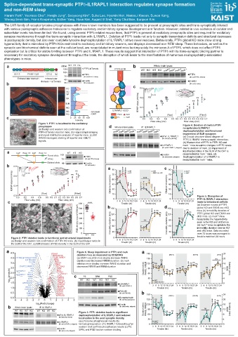
The LAR family of receptor tyrosine phosphatases with three known members has been suggested to be present at presynaptic sites and trans-synaptically interact
with various postsynaptic adhesion molecules to regulate excitatory and inhibitory synapse development and function. However, related in vivo evidence at cellular and
subcellular levels has been limited. We found, using several PTPδ-related mouse lines, that PTPδ is present at excitatory presynaptic sites and required for excitatory
synapse maintenance through the trans-synaptic interaction with IL1RAPL1. Deletion of PTPδ leads not only to synaptic transmission deficits and structural decreases
in postsynaptic density, but also near complete tyrosine dephosphorylation of IL1RAPL1 at two novel residues. Behaviorally, PTPδ global KO mice show strong
hyperactivity that is mimicked by PTPδ KOs restricted to excitatory and inhibitory neurons, and displays decreased non-REM sleep. These behaviors, as well as the
synaptic and biochemical deficits seen at the cellular level, are recapitulated in mutant mice lacking solely the miniexon A of PTPδ, which does not affect PTPδ
expression but is critical for stable binding between PTPδ and IL1RAPL1. These results suggest that interaction of PTPδ with its trans-synaptic binding partner is
necessary for excitatory synapse development throughout the brain, the disruption of which leads to the manifestation of numerous neuropsychiatry-associated
phenotypes in mice.
a (KDa) WT HT Ho a b Whole brain lysate
135 - PTPδ-tdTomato PTPδ-ECD WT KO WT KO meA +/+ meA –/– meA +/+ meA –/–
100 -
IG1-3 FNIII 1-3,8 D1-2 td PTPδ PTPδ
75 - [IB: anti-PTPδ, C-term] 1 [IB: anti-PTPδ, N-term]
Extracellular domain Intracellular 48 - β-actin
b domain + tdTomato 35 - [IB: anti-β-actin] 3 β-actin
[IB: anti-β-actin]
DAPI CC 1 c 0.9
SO tdTomato
SP SO
CC 2
SR SP 0.5 mV *
SLM SR 2 0.6 3 ms **
MO 1 ** *
SG SLM fEPSP slope (mV/ms)
PO *
MO 3 0.3 *
IL1RAPL1-ECD
SG tdTomato
500 PO DAPI FNIII meA [Raw] [Fitted]
c 0 25 50 75 100 IG meB 0.0 0.0 0.2 0.4 0.6 0.8 0.0 0.1 0.2 0.3 0.4 0.5 0.6 0.7
tdTomato Intensity Fiber volley (mV) Fiber volley (mV)
vGluT2
Figure 1. PTPδ is localized to the excitatory
presynapse d Whole brain lysate IP: IL1RAPL1 Figure 4. Deletion of meA in PTPδ
(a) Design and western blot confirmation of +/+ –/– +/+ –/– +/+ –/– +/+ –/– recapitulates IL1RAPL1
PTPδ-tdTomato reporter mice. (b) Hippocampal imaging meA meA meA meA meA meA meA meA dephosphorylation and functional
and signal distribution analysis of reporter mice. (c) EM pTyr, IL1RAPL1 impairment of SLM synapses
DAB/immunogold staining of reporter and vGluT2 [IB: anti-pTyr (4G10)] (a) Crystal structure-based diagram of
signals. PTPδ-IL1RAPL1 trans-synaptic interaction.
(b) Western blot of PTPδ in PTPδ KO and
IL1RAPL1 meA mice reveal no changes in PTPδ levels
–/–
[IB: anti-IL1RAPL1, stripped] due to deletion of meA. (c) Impairment of
a loxP Exon 13 loxP Exon 14 WT HT KO input/output ratios in the SLM of the CA1 is
–/–
Floxed allele 100 β-actin recapitulated in meA mice. (d)
PTPδ [IB: anti-β-actin, stripped] Dephosphorylation of IL1RAPL1 is
HTNC 75 [IB: anti-PTPδ, N-term] recapitulated in meA mice.
–/–
Deleted allele 48 β-actin
b 0.9 [IB: anti-β-actin] a c
** 90 WT cWT 90 meA +/+
fEPSP slope (mV/ms)
0.5 mV 75 KO EMX1-cre; cKO 75 meA –/–
0.6 3 ms ** *
Distance moved (m)
* 60 60
45 Distance moved (m) 45
0.3
30 30
[Raw] [Fitted] 15 15
0.0 Figure 5. Disruption of
0.0 0.2 0.4 0.6 0.8 0.0 0.1 0.2 0.3 0.4 0.5 0.6 0.7 0 0 PTPδ-IL1RAPL1 interaction
c Fiber volley (mV) Fiber volley (mV) 3 6 9 12 15 18 21 24 3 6 9 12 15 18 21 24 3 6 9 12 15 18 21 24 leads to behavioral deficits
PSD density (count/1000mm²) 600
WT KO PSD b Timebin (hr) Timebin (hr) d Timebin (hr) (a) Distance moved of PTPδ
500 150 150 global KO and EMX1-cre cKO
mice (b) Immobility duration of
400 120 120 PTPδ global KO and EMX1-cre
cKO mice. (c) meA mice
–/–
300 recapitulate the hyperactivity
90 90
200 Immobility duration (min) Immobility duration (min) seen in the KO and cKO mice.
–/–
60 60 (d) meA mice recapitulate the
100 immobility duration seen in KO
30 30 and cKO mice. Data recorded
0 over 72 hours was averaged to
WT KO
Figure 2. PTPδ deletion leads to functional and structural impairments 0 0 timebin-matched 24 hours.
(a) Design and western blot confirmation of PTPδ KO mice. (b) Input/Output ratios in 3 6 9 12 15 18 21 24 3 6 9 12 15 18 21 24 3 6 9 12 15 18 21 24
the SLM of the CA1. (c) EM analysis of PSD density in the SLM of the CA1. Timebin (hr) Timebin (hr) Timebin (hr)
a 4.5 Figure 6. Sleep impairment in PTPδ and meA a
Pyk2
ARHGAP26
Src PDHA1 deletion mice as measured by EEG/EMG 180 [WAKE] [NREM] [REM] 24
4.0 PTPRD;PTPRS;PTPRF PCM-1;MBD1 (a) EMX1-cre;cKO mice display increase WAKE 150 ns 20
IGF1R;INSR IRS2 duration and decreased NREM duration. (b) meA
MAG
3.5 CLCN6 Epha4 deletion mice display increase WAKE duration and 120 16
Yes MAG
CKMT2;CKMT1A SLC38A2 decreased NREM and REM duration
3.0 IL1RAPL1 Hck JNK2 Duration (min) WAKE, NREM 90 12 REM Duration (min)
-log 10 (p-vaule) 2.5 IL1RAPL1 TANC2 PCM-1 c WT P2 KO WT KO WT KO 60 8
MAG
Arg
Kv1.1
PDHA1
CPD
SPM
PSD
TGM5
FGFR3
2.0
4
30
SYT7
DDX27
cWT
piccolo
ATP1A3
EMX1-cre; cKO
1.5
ARHGAP27
OPALIN
ACLY CRMP-2 IL1RAPL1 0 3 6 9 12 15 18 21 24 3 6 9 12 15 18 21 24 3 6 9 12 15 18 21 24 0
[IB: anti-IL1RAPL1]
NYAP2
1.0 EHOC-1 PNPLA8 Timebin (hr) Timebin (hr) Timebin (hr)
CASKIN1 HSPE1
KIFC2 PDHA1 PSD-95
0.5 FKBP12 PDHA1 [IB: anti-PSD-95] b
CMTM4 GPRC5B 180 [WAKE] [NREM] [REM] 24
0.0 Synaptophysin
-5 -4 -3 -2 -1 0 1 2 3 4 5 [IB: anti-Synaptophysin] 150 20
log (Fold change) β-actin 120 16
2
b Whole brain lysate IP: IL1RAPL1 [IB: anti-β-actin, stripped] ns
WT KO WT KO WT KO WT KO Duration (min) WAKE, NREM 90 12 REM Duration (min)
Figure 3. PTPδ deletion leads to significant
pTyr, IL1RAPL1 dephosphorylation of IL1RAPL1 and reduced 60 8
[IB: anti-pTyr (4G10)]
localization to the post synaptic density 30 4
IL1RAPL1 (a) pTyrosine phosphoscan results (b) meA +/+
meA
–/–
[IB: anti-IL1RAPL1, stripped] Immunoprecipication of IL1RAPL1 followed by pTyr 0 0
western blot confirms phosphoscan results (c) P2, 3 6 9 12 15 18 21 24 3 6 9 12 15 18 21 24 3 6 9 12 15 18 21 24
β-actin SPM, and PSD fraction western blotting Timebin (hr) Timebin (hr) Timebin (hr)
[IB: anti-β-actin, stripped]