
Page 33 - Q. Neuroscience
P. 33
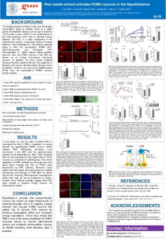
Pine needle extract activates POMC neurons in the Hypothalamus
                                          Lee DK , Kim EA , Byeon EH , Kang D , Han J , Hong SG 1,2
                                                                      1,2
                                                               1
                                                      1
                                                                            1,2
                                               1
                School of Medicine  1 Department of Physiology and Convergence Medical Sciences, School of Medicine, Gyeongsang National University, Jinju, Republic of Korea
                              2 Institute of Health Sciences, School of Medicine, Gyeongsang National University, Jinju, Republic of Korea
                                                                                                     Q-19
        BACKGROUND
   Prolonged excessive energy intake over the energy
   consumption leads to obesity which is a major
   cause of metabolic disease such as type 2 diabetes.
   The Arcuate nucleus (ARC) of the hypothalamus is
   the most important brain area to regulate energy
   balance. The ARC is a major component of the
   melanocortin system, a regulation center of energy
   balance in the hypothalamus. Two distinct neuronal
   types of ARC are anorexigenic POMC (Pro-
   opiomelanocortin)  and  orexigenic  NPY
   (Neuropeptide Y) / AgRP (Agouti related peptide)
   neurons. Also, the POMC neurons in the ARC are
   known  as  an  energy  expenditure  enhancing
   neurons. In addition, the pine needle contains
   strong antioxidant polyphenols and has capability of
   lipolysis and improve elevated blood glucose levels                  Fig 4. Mice orally treated with PNE demonstrated reduced body weight and feeding behaviors.
   by obesity. However, the role of pine needle extract                 (A) Schematic timeline depicting food intake and bodyweight measurement after HFD fed (B)
   (PNE) onto the hypothalamic POMC neurons                             Except for NCD, HFD-fed mice exhibited reduced body weight by daily oral injection of PNE
                                                                        (NCD+Water, n=14; NCD+PNE, n=13; HFD+Water, n=16; HFD+PNE, n=15, respectively) for 2
   remain unclear.                                                      weeks. (C) Body weight, (D) food intake (day and night, n=9), and (E) fat and lean mass (n=6)
                                                                        were measured after HFD fed. Except for lean mass, body weight, food intake, and fat mass
                AIM                                                     were reduced by PNE treatment in HFD fed group. *P<0.05, ***P<0.001, ****P<0.0001 vs. Con.
                                                                        All data are shown as mean ± SEM.
   1. Does PNE regulate hypothalamic areas related in energy   Fig 1. Orally injected PNE increased c-fos expression in both the ARC and PVN as
                                     compared to water injected into the control groups. (A) Schematic illustration of the
                                     hydrothermal PNE extract method. (B) Immunohistochemical staining of the c-fos
   balance regulation?               expression in the ARC and PVN after 200 mg/kg of PNE treatment. (C) and (D)
                                     Quantitative analyses of fluorescent intensity of c-fos expression in the ARC and PVN
   2. Does PNE activate hypothalamic POMC neurons?  after PNE treatments (Con, n=6; PNE 100 mg/kg, n=8; PNE 200 mg/kg, n=9),
                                     respectively. **P<0.01, ****P<0.0001 vs. Con. All data are shown as mean ± SEM
   3. Does PNE regulate feeding behaviors?
   4. Does PNE regulate glucose homeostasis?
   5. Does PNE related in the regulation of energy expenditure
   via BAT thermogenesis?
            METHODS
   Slice preparation and electrophysiological recordings
   Immunohistochemistry (IHC)
   Measurement of body weight, food intake, and body mass
   composition                                                          Fig 5. PNE improved leptin resistance in HFD-induced obese mice. (A) Intraperitoneal glucose
   Intraperitoneal glucose tolerance test (IPGTT)                       tolerance test (IPGTT, 2g/kg) for plasma glucose kinetics after PNE oral injection (NCD+Water,
                                                                        n=8; NCD+PNE, n=10; HFD+Water, n=14; HFD+PNE, n=17, respectively). The mice were
                                                                        fasted overnight prior to the IPGTT. (B) The area under the curve for IPGTT. (C) Changes in
   Western blot Analysis                                                fasting plasma glucose levels in NCD and HFD fed mice. (D) Changes in plasma leptin levels 1
                                                                        hr after PNE treatment. (E) Representative immunoblots for UCP 1 (32 kDa) and β-actin (42
   Blood plasma leptin assay                                            kDa) and quantitative analysis of UCP1 expression levels in the BAT of HFD fed mice after daily
                                                                        PNE treatment for 2 weeks (n=4 per group). *P<0.05, **P<0.01 vs. Con. All data are shown as
                                                                        mean ± SEM. PNE increase UCP1 expression in the iBAT after PNE treatment. Western
            RESULTS                                                     immunoblotting data showing that both NCD and HFD fed mice, PNE increased UCP1
                                                                        expression in the iBAT (NCD+Water, n=4; NCD+PNE, n=4; HFD+Water, n=4; HFD+PNE, n=4).
                                                                        * P<0.05, *** P<0.001 vs. Control. . All data are shown as mean ± SEM
   In this study, we use hot water extracted PNE to
   investigate the role of PNE in regulation of energy
   balance via hypothalamic POMC neurons. Orally
   injected  PNE  (200mg/kg)  increases  c-fos
   expression in the ARC and the paraventricular  Fig 2. PNE depolarized arcuate POMC neurons, but this elevation was inhibited by
                                      the MC4R blockade. (A) Brightfield, fluorescent (eGFP), and a merged image of
   nucleus of the hypothalamus. Also, PNE increased  targeted POMC neuron in the ARC for the patch-clamp recording. (B) Representative
   20% of c-fos expression in the hypothalamic POMC  recording trace showing the PNE effect on POMC neurons. (C) Quantitative analysis
                                      of the PNE effect on POMC neurons. Among the 15 POMC neurons, 10 neurons were
   neurons as compared to control group. And, direct  depolarized after PNE treatment. (D) Representative recording trace showing the
                                      effect of blockade of MC4R after PNE treatment on POMC neurons. (E) Quantitative
   application of PNE (200μM) on the brain slice with  analysis of blockade of MC4R on POMC neurons after PNE treatment. In all of 9
                                      recorded POMC neurons, PNE-evoked depolarization was blocked by SHU-9119 (100
   patch clamp showed that 78% of POMC were  nM). (F) Single effect of SHU-9119 did not change the membrane potential at 100 nM
   depolarized by PNE (200μM). Moreover, body  concentration. ***P<0.001 vs. Con. All data are shown as mean ± SEM. SHU, SHU
                                      9119.
   weight and food intake were decreased by 2 weeks
   consecutive oral injection of PNE after 12 weeks                     Figure 6. Putative mechanism of PNE on melanocortin system. Oral injection of PNE enhances
                                                                        ARC POMC neuronal activity and subsequently activate PVN MC4R neurons. This series of
   high fat fed. Parallelly, PNE improved blood glucose                 activation of the melanocortin system may contribute to enhancement of energy expenditures
                                                                        and reduction of body weight and food intake.
   levels after high fat fed as compared to control.
   Overall, these findings strongly suggest that PNE                           REFERENCES
   plays pivotal to regulate energy balance via
   melanocortin system.                                                1. Romieu I, Dossus L, Barquera S, Blottière HM, Franks PW,
                                                                       Gunter M, et al. Energy balance and obesity: what are the main
         CONCLUSION                                                    drivers? Cancer Causes Control. 2017; 28: 247–258.
                                                                       2. Lumeng CN, Saltiel AR. Inflammatory links between obesity and
   Hypothalamic arcuate and paraventricular                            metabolic disease. J Clin Invest. 2011; 121: 2111–2117.
   nucleus are known as major components of
   melanocortinergic circuit to regulate energy
   balance. And arcuate POMC neurons has                                 ACKNOWLEDGEMENTS
   critical role to activate PVN neurons by                             This research was supported by grants from the Basic Science
   showing anorexigenic effect and increased                           Research Program through the National Research Foundation of
   energy expenditure. These data shows that                           Korea (NRF) funded by the Ministry of Education
                                                                       (2016R1D1A3B03934279) and the Korea Research Institute of
   PNE is one of the strong modulator of POMC                          Bioscience & Biotechnology (KRIBB) Research Initiative Program
   neuronal activity to improve diet-induced  Fig 3. Blockade of MC3/4Rs inhibited PNE-induced activation of POMC neurons,  (KGM4622013).
   obesity and metabolic abnormalities induced  while PNE increased c-fos expression in the arcuate POMC neurons of the
                                      hypothalamus. (A) Images of fluorescence microscopy showing that POMC neurons
   by obesity including heart diseases, type 2  (green) expressed c-fos (red) after oral injection of PNE (200 mg/kg, middle panel).  Contact information
                                      However, PNE-mediated elevation of c-fos expression in the POMC neurons was
   diabetes.                          inhibited by MC4R antagonist SHU-9119 (0.1 mg/kg, i.p.). SHU-9119 was given
                                      intraperitoneally before 30 min to oral injection of PNE. Scale bar: 50 μm. (B)
                                      Quantitative analysis of POMC and c-fos colocalization after single or combination
                                      treatment of PNE and PNE+SHU9119 treatment (Con, n=6; PNE, n=5; PNE+SHU-  Eun A Kim E-mail jbw01151@naver.com
                                      9119, n=7). ****P<0.0001 vs. Con; ####P<0.0001 vs. PNE. All data are shown as
                                      mean ± SEM.                      Correspondence: dklee@gnu.ac.kr (D.K.L.)