
Page 11 - O. Microbiology
P. 11
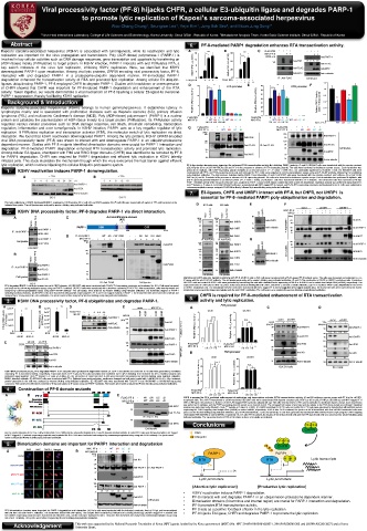
Viral processivity factor (PF-8) hijacks CHFR, a cellular E3-ubiquitin ligase and degrades PARP-1
to promote lytic replication of Kaposi’s sarcoma-associated herpesvirus
Woo-Chang Chung 1 , Seungrae Lee 1 , Yejin Kim 1 , Jong Bok Seo 2 , and Moon Jung Song 1 *
1 Virus-Host Interactions Laboratory, College of Life Sciences and Biotechnology, Korea University, Seoul 02841, Republic of Korea; 2 Metabolome Analysis Team, Korea Basic Science Institute, Seoul 02841, Republic of Korea
15 *** *** RTA promoter 70 *** ** PAN promoter
Abstract 6. PF-8-mediated PARP1 degradation enhances RTA transactivation activity. 60 50
10
*** 40
Kaposi’s sarcoma-associated herpesvirus (KSHV) is associated with tumorigenesis, while its reactivation and lytic A 16 *** *** B 90 *** *** *** C FLAG-RTA - + - + - + - + Relative kRp-Luc activity Relative PAN-Luc activity
MYC-PF-8
+
FLAG-RTA
replication are important for the virus propagation and transmission. Poly (ADP-ribose) polymerase-1 (PARP-1) is 16 12 *** 15 60-Luc activity 90 *** *** *** RTA promoter (kDa) - - + - - + - - 70 + + + + *** ** - + - - - + + + PAN promoter 30
5
+
-
Anti-PARP-1
involved in key cellular activities such as DNA damage responses, gene transcription and apoptosis by transferring an 12 60 MYC-PF-8 120 60 - 1.0 1.1 0.7 0.6 Anti-PARP-1 20
(kDa)
120
Anti-PAR
(ADP-ribose) moiety (PARylation) to target protein. In KSHV infection, PARP-1 interacts with and PARylates RTA, a relative kRp-Luc activity 4 relative kRp-Luc activity 8 relative PAN-Luc activity 90 90 50 Anti-PAR 1.0 1.1 0.7 0.6 Anti-FLAG 10 0
0
+
+
-
-
+
Anti-FLAG -
+
key switch molecule of the virus lytic replication, inhibiting KSHV replication. Here, we identified that KSHV 8 4 10 relative PAN 30 40 Anti-FLAG FLAG-RTA - + WT + ΔN ΔC ΔI WT - ΔN ΔC ΔI - FLAG-RTA - - + WT + ΔN ΔC ΔI WT - ΔN ΔC ΔI -
-
+
+
-
MYC-PF-8 Anti-MYC
downregulated PARP-1 upon reactivation. Among viral lytic proteins, ORF59 encoding viral processivity factor, PF-8, Relative kRp-Luc activity 30 - - Relative PAN-Luc activity 30 Anti-FLAG Anti-MYC 1.00 - 1.51 0.95 1.53 0.88 MYC-PF-8 1.00 - 1.45 1.15 1.48 1.01
Anti-MYC
Anti-α-tubulin
interacted with and degraded PARP-1 in a proteasome-ubiquitin dependent manner. PF-8-mediated PARP-1 0 - + + + - 5 FLAG-RTA 0 - + + + - 20 Anti-MYC Anti-α-tubulin PARP-1 enzymatic activity
FLAG-RTA
IP : Anti-FLAG
Cell lysates
degradation enhanced the transactivation activity of RTA and promoted lytic replication. Among cellular E3 ubiquitin- FLAG-RTA 0 - + + + - FLAG-RTA 0 - + + + - ++ ++ IP : Anti-FLAG 10 Cell lysates FLAG-RTA - - + - WT + ΔN + ΔC + ΔI +
- +
-
MYC-PF-8
***
RTA promoter
ligases ubiquitinating PARP-1, PF-8 employed CHFR to degrade PARP-1. Studies with knockdown or overexpression D 70 *** *** 15 ** 0 *** -*** - + ++ ++ ** 70 0 *** ** F PAN promoter (kDa) 120
1.2
15
PAN promoter RTA promoter
E
***
+
+
+
+
of CHFR showed that CHFR was important for PF-8-induced PARP-1 degradation and enhancement of the RTA 15 60 *** FLAG-RTA RTA promoter + + - - 70 - *** FLAG-RTA 60 - PAN promoter + + - - - 1.0 - Anti-PAR
+
+
-
ΔN ΔC ΔI WT
activity. Taken together, our results demonstrate a viral mechanism of PF-8 hijacking a cellular E3-ligase to overcome 50 MYC-PF-8 10 - 1.00 - WT ΔN ΔC ΔI WT ΔN ΔC 60 ΔI MYC-PF-8 50 - 1.00 - WT 1.45 1.15 1.48 1.01 ΔN ΔC ΔI 120 90
1.51 0.95 1.53 0.88
10
PARP-1 suppression, thereby facilitating KSHV replication. Relative kRp-Luc activity 10 Relative PAN-Luc activity 40 Relative kRp-Luc activity PARP-1 enzymatic activity 50 40 Relative PAN-Luc activity 40 ΔI Absorbance) 0.8 ** ** 90 Anti-FLAG IP: Anti-FLAG
Background & introduction 5 Relative kRp-Luc activity 30 20 5 1.2 Relative PAN-Luc activity - 30 FLAG-RTA 30 - - + - WT + ΔN + ΔC + Relative PARP-1 activity + 0.6 75 Anti-MYC
MYC-PF-8
Kaposi’s sarcoma-associated herpesivirus (KSHV) belongs to human gammaherpesvirus. It establishes latency in 5 10 20 (kDa) 20 10 (450 nm 0.4 54
120
lymphocytes mainly and is associated with proliferative diseases such as Kaposi’s sarcoma (KS), primary effusion 0 1.0 0 10 90 0 Anti-PAR 120 90 Anti-PARP-1
0
-
+
+
-
-
-
+
+
0 -
+
+
-
+
+
- +
lymphoma (PEL) and multicentric Castleman’s disease (MCD). Poly (ADP-ribose) polymerase-1 - - (PARP1) is a nuclear - - FLAG-RTA MYC-PF-8 + -MYC-PF-8 +A + - - - + + - + - WT - + - + - ΔN ΔC ΔI - + - ΔN ΔC ΔI + MYC-PF-8 120 - - - + - - WT + - ΔN ΔC ΔI WT - ΔN ΔC ΔI - 120
FLAG-RTA
FLAG-RTA
+ty
FLAG-RTA
- 0.2
-FLAG-RT
+ Absorbance)
0
ΔN ΔC ΔI WT
WT
MYC-PF-8
-
+ ΔN ΔC ΔI WTΔN ΔC ΔI WT
ΔN ΔC ΔI
-
-
-
+
+
+
protein and catalyzes the polymerization of ADP-ribose moiety to a target protein (PARylation). Its 1.00 PARylation MYC-PF-8 - - + - WT ΔN ΔC ΔI WT 1.011.00 + WT 0.8 + ΔN ΔC ΔI 1.53 0.88 ** FLAG-RTA - - + - WT ΔN ΔC ΔI WT 1.00 ΔN ΔC ΔI 1.48 1.01 Anti-FLAG IP: Anti-FLAG 90 Anti-FLAG
1.51 0.95 1.53 0.88 activity
1.45 1.15
1.45 1.15 1.481.00
1.51 0.95
MYC-PF-8
**
0
90
regulates various cellular processes such as DNA damage response, cell death, chromatin remodeling, transcription 1.00 1.51 0.95 1.53 0.88 0.6 1.00 1.45 1.15 1.48 1.01 MYC-PF-8 - WT ΔN ΔC ΔI 75 Cell lysates
75
PARP-1 enzymatic activity
+
+
regulation, inflammation and even tumorigenesis. In KSHV infection, PARP1 acts as a key negative regulator of lytic G FLAG-RTA - Relative PARP-1 activi + + PARP-1 enzymatic activity FLAG-RTA - + + + + + Anti-MYC 54 Anti-MYC
PARP-1 enzymatic activity ΔC--
+ 54
replication. It PARylates replication and transcription activator (RTA), the molecular switch of 1.2 lytic replication via direct MYC-PF-8 (450 nm WT 1.2 0.4 ΔN ΔI FLAG-RTA - - + - WT + MYC-PF-8 ΔC +- +- ΔI WT ΔN ΔC ΔI 54 Anti-α-tubulin
MYC-PF-8
(kDa)
ΔN
(kDa)
interaction. We found that KSHV reactivation downregulated PARP1. Among the lytic proteins, KSHV ORF59-encoded 1.2 120 Anti-PAR (kDa) 120 120 120 90 Anti-PARP-1
Anti-PAR
(450 nm Absorbance) in an ubiquitin-proteasome
viral DNA processivity factor (PF-8) was shown to interact with and downregulate PARP-1 1.0 1.0 90 1.0 0.2 120 90 Anti-PAR
Relative PARP-1 activity
dependent manner. Studies with PF-8 mutants identified dimerization domains were crucial for 0.8 PARP-1 interaction and 120 0.8 Anti-FLAG 120 IP: Anti-FLAG 120 90 Anti-FLAG IP: Anti-FLAG
90
Anti-FLAG
**
**
degradation. PF-8-mediated PARP1 degradation enhanced RTA transactivation activity and promoted **lytic replication. 0.8 90 MYC-PF-8 ** 0 - ** WT ΔN ΔC ΔI 75 90 Anti-FLAG IP: Anti-FLAG Cell lysates
0.6 to be recruited by PF-8
Among cellular E3-ligases, checkpoint with FHA and RING finger domains (CHFR) was identified 75 ** Relative PARP-1 activity (450 nm Absorbance) 0.6 90 75 Anti-MYC
for PARP1 degradation. CHFR was required for PARP1 degradation and efficient lytic replication in KSHV latently Relative PARP-1 activity (450 nm Absorbance) 0.6 54 Anti-MYC 75 54 54 Anti-MYC Anti-MYC
0.4
infected cells. This study elucidates the mechanism through which the virus overcomes the host barrier against efficient 120 0.4 54 120 54 Anti-α-tubulin
Anti-PARP-1
0.4
Anti-PARP-1
lytic replication, which involves hijacking the cellular Ubiquitin-proteasome system. PF-8 dimerization domains were important for enhanced RTA transactivation 120 activity by inhibiting PARP-1 activity. (A and B) HEK293T cells were transfected with the reporter construct
90
90
Anti-PARP-1
0.2 pGL3-kRp-luc (A) or 120 pGL3-PAN RRE-luc (B) and PF-8 in the presence or absenc 90e of RTA-expressing plasmid. The cells were harvested for luciferase reporter assays. Each transfection was
0.2
120
1. KSHV reactivation induces PARP-1 donwregulation. 0 performed in 0.2 triplicate, with EGFP-expressing plasmid included as an internal control (***, P value < 0.005; N.S., not statistically significant by Student’s t test). (C) HEK293T cells were
Anti-FLAG
90
90
Anti-FLAG 48 h.
120 Cells were subjected to coimmunoprecipitation assays using anti-FLAG-M2 antibody, followed by immunoblotting
cotransfected with PF-8- and RTA-expressing constructs and incubated for
0
ΔN PARP-1 band intensities. (D
Anti-FLAG with the reporter construct pGL3-kRp-luc (D) or pGL3-PAN
75
MYC-PF-8
-
A B MYC-PF-8 - WT ΔN ΔC ΔI using indicated antibodies. The digit numbers indicates WT relative ΔC ΔI 90 Cell lysates and E) HEK293T cells were transfected Cell lysates
75
0
ΔN presence or absence of RTA-expressing plasmid. The cells were harvested for luciferase reporter assays. Each transfection was performed in triplicate, with
RRE-luc (E) and PF-8
WT in the
- mutants
ΔC
MYC-PF-8
Cell lysates
ΔI
Anti-MYC
Anti-MYC 0.005).
EGFP-expressing plasmid included as an internal control (***, P value < 75 (F) HEK293T cells were transfected with MYC-PF-8 mutants. The transfected cells were harvested at 48 hpt.
Anti-MYC
54
PARP-1 inhibition activity of the 50 μg of cell lysates was analyzed by PARP-1 assay kit with histone-coated 54 strip wells at 450 nm absorbance. Statistical analysis was performed using Student’s t
test (** denotes a P value of <0.01).(G) HEK293T cells were cotransfected with MY 54C-tagged PF-8 mutants and FLAG RTA-expressing constructs and incubated for 48 h. Cells were subjected to
54
54
Anti-α-tubulin
coimmunoprecipitation assays using anti-FLAG-M2 antibody, followed by immunoblotting using indicated antibodies. Anti-α-tubulin
54 Anti-α-tubulin
7. E3-ligases, CHFR and UHRF1 interact with PF-8, but CHFR, not UHRF1 is
essential for PF-8–mediated PARP1 poly-ubiquitination and degradation.
FLAG-RNF144a
The lytic replication of KSHV decreased PARP-1 expression. KSHV-positive BC-3 cells (A) and KSHV-negative DG-75 cells (B) were treated with 20 ng/mL of TPA and harvested at the D E F
indicated time points. The cell lysates were analyzed by western blotting using indicated antibodies. BC-3 cells SLK cells shCtrl shCHFR shUHRF1
2. KSHV DNA processivity factor, PF-8 degrades PARP-1 via direct interaction. FLAG-RNF146 FLAG-CHFR FLAG-UHRF1 shCHFR MYC-PF-8 - - - + + - + + - - - + + - + + - - - + + - + +
HA-Ub
Automodification domain - - Vector FLAG-PF-8 Vector FLAG-PF-8 shCtrl (kDa)
A C Automodification domain MYC-PF-8 - + + + + + (kDa) 120 (kDa) 90 Anti-CHFR 120 Anti-HA
(AD)
(AD)
(kDa) 120
120
Vector FLAG-PF-8 PARP-1 DBD BRCT CAT 75 90 Anti-PARP-1 90 90 90 90 Anti-UHRF1 75 90
PARP-1 1 DBD BRCT 524 CAT Anti-MYC 54 Anti-α-tubulin IP: Anti-PARP-1 120 Anti-PARP-1
Anti-PARP-1 1 372 372 524 1014 75 75 Anti-CHFR 90
1014
IP : Anti-PARP-1 120 54 54 75
Anti-FLAG
D FLAG-PARP-1 - - WT AD CAT DBD - - WT AD 90 CAT DBD Anti-FLAG Anti-MYC
+
+
+
MYC-PF-8 -
MYC-PF-8
Anti-PARP-1 FLAG-PARP-1 - + - + WT AD CAT + DBD - - + - WT + AD 75 + CAT + DBD Anti-FLAG IP: Anti-FLAG IP: Anti-FLAG G
Cell lysates (kDa) (kDa) - + + + + + - + + + + + 120 120
75
Anti-FLAG 75 54 Anti-MYC 120 90 90 Anti-PARP-1 shUHRF1 90 Anti-PARP-1
Anti-MYC shCtrl 75
HEK293T IP: Anti-FLAG 120 120
120 120 90 90 Anti-UHRF1 Cell lysates Anti-MYC
B FLAG-PF-8 90 120 90 90 75 Anti-PARP-1 90 90 90 Anti-UHRF1
Vector Anti-MYC 75 54 75 54 Anti-CHFR 54 Anti-α-tubulin 54 Anti-α-tubulin
Anti-PARP-1 54 54 120 Anti-FLAG Anti-FLAG
Anti-FLAG
IP : Anti-PARP-1 90 Anti-FLAG shCtrl shCHFR
Anti-FLAG 75 54 54 Anti-α-tubulin DOX - + - +
43 43 (kDa)120
Anti-PARP-1 35 35 54 Cell lysates Cell lysates 90 Anti-PARP-1
Cell lysates Identification of E3 ubiquitin ligases interacting with PF-8. (A) BC-3 cells or SLK cells were transduced with a FLAG-tagged PF-8 lentiviral vector. The cells were harvested and subjected to a co-
Anti-FLAG 24 1.0 0.6 knockdown 1.3 cells. Knockdown
1.2
24 54 IP assay with the anti-FLAG-M2 antibody. The cell lysates were analyzed by western blotting using indicated antibodies. (B and C) The construction of CHFR or UHRF1
BC-3 IP: Anti-FLAG Cell lysates Anti-α-tubulin HEK293T cells and control HEK293T cells were generated by transducing the cells with a lentiviral vector expressing shCHFR or shUHRF1 and a control shor 90t hairpin RNA (shRNA), respectively. The
IP: Anti-FLAG
Anti-CHFR
Cell lysates
75 PARP1 poly-ubiquitination in the CHFR
PF-8 degraded PARP-1 via direct interaction with CENT domain. (A) HEK293T cells were transfected with FLAG-PF-8 expressing constructs and incubated for 48 h. Cells were harvested expression levels of CHFR (B) or UHRF1 (C) were analyzed by western blotting with anti-CHFR, anti-UHRF1, and anti-α-tubulin antibodies. (D) PF-8–mediated
Cell lysates
and analyzed by coimmunoprecipitation assay using anti-PARP-1 antibody. (B) BC-3 cells were transduced with a lentivirus expressing FLAG-PF-8. After transduction, cells were harvested and or UHRF1 knockdown cells. The knockdown HEK293T cells were transfected with MYC-tagged PF-8 and hemagglutinin (HA)-tagged ubiquitin (Ub). The transfected cells were harvested at 48 h post-
120
analyzed by coimmunoprecipitation assay using anti-FLAG-M2 antibody. The cell lysates were analyzed by western blotting using indicated antibodies. (C) Schematic diagram of PARP-1 transfection and assayed by immunoprecipitation with the anti-PARP1 antibodies. The cell lysates were analyzed by western blotting using indicated antibodies. Anti-RTA
indicating functional domains. (D) HEK293T cells were transfected with plasmids expressing MYC-PF-8 and FLAG-tagged mutants of PARP-1. The transfected cells were harvested at 48 hpt and
90
analyzed by IP assay using anti-FLAG antibodies. The lysates were further analyzed by Western blotting using using indicated antibodies. 8. CHFR is required for PF-8–mediated enhancement of RTA transactivation 0.3
1.0
35
3. KSHV DNA processivity factor, PF-8 ubiquitinates and degrades PARP-1. A activity and lytic replication. Anti-K8
RTA promoter
MYC-PF-8
A 1.2x10³ *** B 6x10⁴ *** D FLAG-PF-8 - sh Ctrl - + + ++ ++ E vector vector K48R K63R MYC-PF-8 K48R K63R A A 8 8 shCtrl shCtrl RTA promoter 8 8 shCHFR C D 54 1.0 0.5 Anti-α-tubulin
sh PF8
shCHFR
-
DOX
+
-
PF-8 mRNA lelvels (PF-8/Actin) 9x10² RTA mRNA lelvels (RTA/Actin) 4x10⁴ FLAG-PF-8 1.0 - MG132 + 0.8 - - 1.0 + ++ ++- 0.3 Anti-PARP-1 (kDa) 130 (kDa) HA-Ub 90 - - K48R K63R - - K48R K63R Anti-HA 6 Relative kRp-Luc activity 6 4 *** *** *** *** 6 Relative kRp-Luc activity 6 4 N.S. N.S. (kDa)120 shCtrl + shCHFR + (kDa)120 shCtrl + shCHFR +
5x10⁴
-
+
+
Anti-PARP-1
iSLK.219 cells
0.9
0.9
+
-
MG132
0.4 -
1.0 -
HA-Ub 130
Anti-PARP-1
Anti-HA
Anti-RTA
3x10⁴
6x10²
0.6
0.8
0.6
DOX
-
-
-
-
TPA
0.6
1.0
0.3
0.6
0.6
90
Anti-FLAG
2x10⁴
Anti-K8
3x10²
130
Anti-PARP-1
Anti-α-tubulin
0.7
Anti-α-tubulin
0 1x10⁴ 0 1.0 Anti-FLAG 130 90 90 75 Anti-PARP-1 IP: Anti-PARP-1 IP: Anti-PARP-1 Relative kRp-Luc activity 4 2 Relative kRp-Luc activity 4 2 90 1.0 0.6 1.2 1.3 Anti-PARP-1 90 Anti-PARP-1
DOX - + - + DOX - + - + Anti-α-tubulin 75 90 90 1.0 0.5 0.9 0.9
shCtrl shPF-8 shCtrl shPF-8 Anti-FLAG 2 2 75 Anti-CHFR 75 Anti-CHFR
Anti-FLAG
*** 130 FLAG-RTA 0 - + + + - FLAG-RTA 0 - + + + - 120 120
Anti-PARP-1
+ ++ ++
1.2x10³ *** 6x10⁴ C DOX sh Ctrl + sh PF8 + Anti-PARP-1 130 90 90 75 1.0 1.2 1.1 0.6 0.8 0.6 Anti-PARP-1 FLAG-RTA 0 - 1.00 1.22 1.46 + ++ ++ FLAG-RTA 0 - 1.00 0.97 1.00 + 90 1.0 0.3 Anti-RTA 90 Anti-RTA
MYC-PF-8 -
MYC-PF-8 -
1.00 1.22 1.46 -
- +
+
- +
- +
1.00 0.97 1.00 -
- +
-
-
PF-8 mRNA lelvels (PF-8/Actin) 9x10² RTA mRNA lelvels (RTA/Actin) 4x10⁴ 1.0 0.4 1.0 0.9 0.9 0.6 Anti-RTA 75 1.0 54 1.2 1.1 0.6 0.8 0.6 Anti-FLAG Cell lysate Cell lysate B B B 40 shCtrl PAN promoter 40 shCHFR 35 1.0 0.5 Anti-K8 35 1.0 1.0 0.1 0.1 Anti-K8
MYC-PF-8
+ ++ ++
MYC-PF-8
-
5x10⁴
+ ++ ++
-
PAN promoter
Anti-FLAG
6x10²
3x10⁴
2x10⁴
Anti-α-tubulin
3x10²
0 1x10⁴ 0 1.0 0.7 Anti-K8 54 Anti-α-tubulin 40 30 shCtrl *** *** *** 40 30 shCHFR N.S. 54 Anti-α-tubulin 54 Anti-α-tubulin
DOX - + - + DOX - + - + Anti-α-tubulin *** N.S. iSLK.219 cells BC-3 cells
shCtrl shPF-8 shCtrl shPF-8 30 Relative PAN-Luc activity 30 Relative PAN-Luc activity shCtrl shCHFR
KSHV DNA processvity factor, PF-8 degraded PARP-1 in an ubiquitination-proteasome dependent manner. (A-C) PF-8 knockdown and control iSLK.219 cells were generated by a lentivirus
expressing PF-8 and control shRNA, respectively. Expression levels of PF-8 (A) and RTA (B) transcripts were confirmed by RT-qPCR following DOX treatment for 48 h. Statistical analysis was 20 20 TPA - + - +
performed using Student’s t test (*** denotes a P value of <0.005). (C) The cell lysates of shCtrl and shPF-8 cells were harvested and analyzed by Western blotting using indicated antibodies. Relative PAN-Luc activity 20 Relative PAN-Luc activity 20 (kDa)120
DMSO, dimethyl sulfoxide. (D) HEK293T cells were transfected with PF-8. Eighteen hours posttransfection, media were changed and cells were treated with 1 μM MG132 for 6 h. After treatment, Anti-PARP-1
protein expression in the cells was analyzed by western blotting using indicated antibodies. (E) HEK293T cells were transfected with FLAG-PF-8 and HA-UbK48R or HA-UbK63R-expressing 10 10 90
constructs. The transfected cells were harvested at 48 hpt and subject to IP assay using anti-PARP-1 antibody. The lysates were further analyzed by Western blotting using indicated antibodies. 10 10 90 1.0 0.5 0.9 0.9
4. Construction of PF-8 domain mutants FLAG-RTA 0 - + + + - FLAG-RTA 0 - + + + - 75 Anti-CHFR
+ ++ ++
+ ++ ++
MYC-PF-8 -
+
1.00 1.14 1.25 -
- +
- +
MYC-PF-8 -
- +
+
A B C FLAG-RTA 0 - PF-8–mediated FLAG-RTA 0 - - + - 1.00 0.98 1.03 - 120 Anti-RTA
-
+ ++ ++
MYC-PF-8
FLAG-PF-8 - WT ΔN ΔC ΔI CHFR is required for 1.00 1.14 1.25 MYC-PF-8 1.00 0.98 1.03 90
+ ++ ++ enhancement of replication and transcription activator (RTA) transactivation activity. (A and B) Luciferase reporter assays with PF-8 in the shCHFR-
1.0
MYC-PF-8 - WT ΔN ΔC ΔI transfected cells. The shCHFR-transfected or shCtrl-transfected HEK293T cells were cotransfected with reporter construct 0.1 pGL3-kRP-Luc (A) or pGL3-PAN-Luc (B) (300 ng) and MYC-tagged PF-8
kDa 75 (150 or 300 ng) in the presence or absence of the FLAG-tagged RTA expression plasmid (25 ng). 35 The cells were harvested at 48 h post-transfection for luciferase reporter assays. Each transfection
Anti-K8 activity relative to the RTA alone sample are indicated. Statistical
Anti-MYC was performed in triplicate, and the EGFP-expressing plasmid served as an internal control. The increased fold values of promoter
analysis was performed using Student’s t test (*** denotes a P value of <0.005). (C) CHFR knockdown iSLK.219 cells and control iSLK.219 cells were generated by transduction with lentiviral vectors
0.1
54 expressing the CHFR-targeting short hairpin RNA (shRNA) or control shRNA, respectively. At 48 h after DOX 1.0 treatment, the lysates of shCtrl-transfected cells and shCHFR-transfected cells were
Anti-FLAG processed for western blotting using indicated antibodies. (D) CHFR knockdown BC-3 cells and control BC-3 cells were generated by transduction with lentiviral vectors expressing the CHFR-targeting
Anti-α-tubulin
54 of shCtrl-transfected cells and shCHFR-transfected cells were processed for western blotting using
IP: Anti-FLAG short hairpin RNA (shRNA) or control shRNA, respectively. At 24 h after TPA treatment, the lysates
indicated antibodies. The expression levels of RTA or K8 relative to those of α-tubulin are indicated.
75 BC-3 cells
Conclusions PF-8 PF-8
Anti-MYC
Cell lysate CHFR
(A) The crystal structure of PF-8 as a dimer form (Baltz et al., 2009). (B)The schematic diagram of PF-8 and its domain deletion mutants. (C) HEK293T cells were transfected with FLAG-tagged PAR
or MYC-tagged PF-8 mutants expressing constructs and incubated for 48 h. Cells were harvested and analyzed by coimmunoprecipitation assay using anti-FLAG antibody. The lysates were
further analyzed by Western blotting using indicated antibodies. Ub Ubiquitin
PF-8 PF-8
5. Dimerization domains are important for PARP1 interaction and degradation Ub CHFR
MYC-PF-8 - - WT ΔN ΔC ΔI Ub
A DAPI MYC PARP-1 Merged B HA-UbK63R - + + + + + Ub
(kDa) PARP1 PARP1
Vector 120
90 Anti-HA RTA RTA PF-8 PF-8 Lytic transcripts
MYC-PF-8WT 120
90 Anti-PARP-1 IP: Anti-PARP-1
75
MYC-PF-8ΔN
Anti-MYC
54 Lytic promoters Lytic promoters
MYC-PF-8ΔC 120
90 Anti-PARP-1 [Abortive lytic replication] [Productive lytic replication]
1.0 0.9 0.1 1.3 0.1 1.0
75 • KSHV reactivation induces PARP-1 degradation.
MYC-PF-8ΔI
Anti-MYC • PF-8 interacts with and degrades PARP-1 in an ubiquitination-proteasome dependent manner.
• Dimerization domains (N-terminus and internal region) are crucial for PARP-1 interaction and degradation.
54 Cell lysates
54 Anti-α-tubulin • PF-8 promotes RTA transactivation activity.
• PF-8 acts as a positive feedback effector in the lytic replication.
PF-8 dimerization domains were important for PARP-1 degradation and interaction. (A) HeLa cells were transfected with the indicated constructs, fixed at 24 hpt, and immunostained
with anti-MYC and anti-PARP-1 antibodies. The nuclei were stained with DAPI (blue). The samples were examined by confocal laser scanning microscopy. (B) MYC-tagged PF-8 mutants and • PF-8 hijacks E3-ligase, CHFR and degrades PARP-1 to promote the lytic replication.
HA-UbK63R-expressing constructs were transfected into HEK293T cells, and the cells were incubated for 48 h. They were then harvested and analyzed by coimmunoprecipitation assay using
anti-PARP-1 antibody. The lysates were further analyzed by Western blotting using indicated antibodies.
Acknowledgement This work was supported by the National Research Foundation of Korea (NRF) grants funded by the Korea government (MSIT) (No. NRF-2019R1A6A3A01093571, 2018R1A2B6001363 and 2020R1A2C2013827) and a Korea
University Grant.