
Page 1 - K. Development and regeneration
P. 1
E xpression pattern and putative func tion of E R -stress molec ules
in tooth formation
1
1
1
1
Yam Prasad Aryal , Tae-Young K im , E ui-S eon Lee , E lina Pokharel , S hijin S ung , J ae-K wang J ung , Hitoshi Yamamoto , 2
1
1
C hang-Hyeon An , Wern-J oo S ohn , J ae-Young K im 1#
3
1
2
1 School of Dentistry, IHBR, Kyungpook National University, Daegu , Korea, Department of Histology and Developmental Biology, Tokyo Dental
3
College, Tokyo, Japan, Division of Biotechnology and Convergence, Daegu Haany University, Gyeongsan Korea
BACKGROUND AIM
Endoplasmic reticulum (ER) is the site for protein
folding and modification, but with the accumulation of Although few studies described the role of ER-
unfolded and misfolded proteins in the ER lumen, there is stress molecules, however, they could not address
creation of unfolded protein response (UPR) which in turn whether all ER-stress molecules involved during tooth
activates ER-stress signaling molecules (Ire1, Atf6 and Perk) development or not. Here, the roles of ER-stress
to maintain homeostasis. ER-stress pathway and its relation molecules: Atf6, Ire1, Perk and Xbp1 were elucidated in
has been described in development of various tissues and the cap, bell and secretory stages of tooth morphogenesis
disease model system. Tooth, a type of hard tissue, also through gene expression analysis.
involves ER-stress pathway especially during ameloblast and
odontoblast differentiation (Brookes et al., 2017; Kim et al.,
2014) however, data on the involvement of major ER-stress
sensors in tooth development are lacking.
METHODS
Animal: Mouse embryos were obtained from time-mated pregnant mice that were maintained in an optimal environment. The day that the vaginal plug
was confirmed was designated as embryonic day 0 (E0). Embryos at E14, E16 and postnatal (PN) day 0 were used as representatives of the cap, bell, and
secretory stages of tooth development, respectively.
Quantitative PCR (qPCR) and In situ hybridization: RNA was extracted from the molar tooth germ of E14, E16 and postnatal day 0 mice, and cDNA
was synthesized for qPCR analysis and section in situ hybridization were performed at 68 °C using digoxigenin (DIG)-labeled RNA probes and standard
protocols, as described previously (Aryal et al., 2019).
Histology and Immunohistochemistry: Hematoxylin and eosin (H&E) staining and immunohistochemistry were performed as previously described
(Aryal et al., 2019). Primary antibodies were directed against Ire1, Atf6, Xbp1 and GRP78.
RESULTS
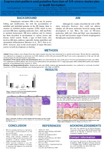
Figure 1. Expression of ER-stress related signaling molecules
in different stages of molar development
Figure 3. Expression patterns of ER-stress related signaling molecules during bell (left)
and secretory stage (right) of tooth development. H&E, Hematoxylin and Eosin staining;
IEE, inner enamel epithelium; OEE, outer enamel epithelium; DP, dental papilla; Od,
odontoblasts; Ab, ameloblasts; SR, stellate reticulum. Scale bars: 50 μm (a–i).
Figure 2. Expression of ER-stress related signaling molecules during cap stage of tooth
development. H&E, Hematoxylin and Eosin staining; EK, enamel knot; DP, dental papilla. Figure 4. Localization patterns of ER-stress related molecules during secretory stage
Scale bars: 50 μm (a–i). of mice molar development. Scale bars: 200 μm (a–f); 50 μm (a’a”-f’f”).
CONCLUSION REFERENCES ACKNOWLEDGEMENTS
This study was supported by the National Research
• ER-stress signaling molecules are expressed at • Adams et al., 2019; Foundation of Korea (grant NRF-2018R1A2A3075600)
various stages of tooth development: cap, bell https://doi.org/10.3389/fmolb.2019.00011 funded by the Ministry of Education, Science and
and secretory stages • Aryal et al., 2019; Technology, Republic of Korea.
• Distinct localization patterns of ER-stress https://doi.org/10.1002/jcp.28635
sensors along secretory odontoblasts and • Brookes et al., 2017; Contact information
ameloblasts suggested their involvement in the https://doi.org/10.3389/fphys.2017.00653
modulation of protein overload especially • Kaneko et al., 2017;
during extracellular matrix secretion https://doi.org/10.1248/bpb.b17-00342 Jae-Young Kim, Department of Biochemistry, School
• Precise functional evaluations of these ER- • Kim et al., 2014; of Dentistry, IHBR, Kyungpook National University,
stress related signaling molecules is necessary https://doi.org/10.1177/0022034514525199 2177 Dalgubeol-daero, Joong-gu, Daegu 41940, Korea
to understand and regenerate the dental hard • Uchibe et al., 2012 Tel: +82-53-420-4998; Fax: +82-53-421-4276
tissue with proper https://doi.org/10.1002/dvdy.23808 E-mail: jykim91@knu.ac.kr