
Page 27 - H. Cell signaling
P. 27
CXXC5, a target for the longitudinal bone growth, regulates growth plate senescence
Sehee Choi, Dasung Lee, Eunhwan Kim, Minguen Yoon, Yeong Chan Ryu and Kang-Yell Choi (Yonsei University)
BACKGROUND AIM
Chondrocytes in cartilage layer proliferate and undergo hypertrophic differentiation and go Approaching a potential therapeutic strategy using
through the remodeling of bone tissue resulting in bone elongation. Currently, many
children undergo early pubertal development with growth plate senescence. These KY19382, a small molecular targeting CXXC5, for
phenomena, known as precocious puberty, is due to the premature termination of
longitudinal bone growth, resulting in short height at adulthood. treatment of children with growth retardation due to
In recent years, Wnt/β-catenin signaling has emerged as a key player in growth plate early growth plate senescence.
maturation. Moreover, mutation of genes involved in the regulation of Wnt/β-catenin
signaling often resulted in impaired bone growth.
In this study, we found that CXXC finger protein 5 (CXXC5), a negative regulator of Wnt/β-
catenin signaling functioning via interaction with the PDZ domain of dishevelled (DVL) in
the cytosol, progressively increased in the resting, proliferative, and hypertrophic
chondrocytes undergoing growth plate senescence. We also found that estrogen, a sex
hormone that is elevated during the pubertal period, induced CXXC5 expression followed
by decrement of β-catenin in chondrocytes.
METHODS
Cells & Animals
For E2 (17β-estradiol; Sigma-Aldrich) treatment, the cells were cultured in phenol red–free DMEM/F12 with 5% charcoalstripped FBS for 24 h followed by serum-free medium for 24 h before the experiment. In case of animals, to manipulate growth plate senescence by
estrogen, 3-wk-old Cxxc5+/+ and Cxxc5−/− male mice received weekly i.m. injections of either 70 μg/kg estradiol (E2) cypionate (Sigma-Aldrich) or vehicle (cottonseed oil) for 3 wk. KY19382 (0.1 mg/kg) was administered daily by i.p. injection to 3- and 7-wk-old mice for
2 wk or to 3-wk-old mice for 10 wk.
Histochemical analyses
The tissues were fixed in 4% PFA, decalcified in 10% EDTA (pH 7.4), dehydrated, embedded in paraffin, and sectioned to 4-μm thickness. The tissues sections were rehydrated and used for further analyses including H&E, TRAP, and IHC staining. The sections were
incubated at 4°C overnight with the following primary antibodies: anti–β-catenin, anti-CXXC5, anti-BrdU, anti-COL2A1, anti-Ki67, and anti-RUNX2. Then, the sections were incubated at room temperature for 1 h with biotinylated anti-mouse or biotinylated anti-rabbit
secondary antibodies. The sections were then incubated in avidin–biotin complex solutions, stained with a DAB kit for 3–30 min, and counterstained with methyl green. For fluorescence staining, the sections were incubated with primary antibody at 4°C overnight,
followed by incubation with anti-mouse Alexa Fluor 488 or anti-rabbit Alex Fluor 555 secondary antibodies at room temperature for 1 hr.
Immunocytochemistry
ATDC5 or C28/I2 cells were seeded on glass coverslip in 12-well culture plates. The cells were incubated with primary antibodies specific for β-catenin (1:100) or CXXC5 (1:200) at 4°C overnight. The cells were washed in PBS and incubated with Alexa Fluor 488 or
Alexa Fluor 555 secondary antibodies (1:200) at room temperature for 1 h. Cell nuclei were counterstained with DAPI for 10 min.
Immunoblot analyses
Immunoblotting was performed with the following primary antibodies: anti–β-catenin, anti-CXXC5, anti-Myc tag, anti-FLAG, anti-p-GSK3α/β, anti-COL2A1, anti-RUNX2, anti-COL10A1, anti-MMP13, anti-ERK and anti–α-tubulin. The samples were then incubated with
horseradish peroxidase–conjugated anti-mouse, anti-rabbit or anti-goat secondary antibodies. Protein bands were visualized with ECL.
Reporter assay
HEK293-TOP cells were seeded into each well of a 24-well plate. The cells were treated with individual compounds at indicated concentration and cultured for 18 h. The cells were then harvested and 20 μl of the supernatant was used to measure luciferase activity.
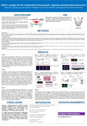
RESULTS
Figure 1 Figure 1. Changes in Wnt/β-catenin pathway and Figure 2. The effects of estrogen on CXXC5
CXXC5 expression during growth plate senescence. expression and the mediation of growth plate
We found that the signatures of Wnt/β-catenin signaling-activated genes senescence.
were significantly down-regulated in the growth plates of the 12-wk-old rats
(Fig 1A). The mRNA level of Cxxc5 was gradually elevated during pubertal
progression (Fig 1B). Immunoblot analyses also showed that CXXC5
gradually increased with the decrement of β-catenin and chondrogenic
markers including COL2A1, RUNX2, COL10A1, and MMP13 in the growth
plates of mice undergoing pubertal progression (Fig 1D). The inverse
correlation between CXXC5 and Wnt/β-catenin signaling was verified by
immunohistochemical (IHC) analyses in all growth plate zones of 3 to 12-
wk-old mice (Fig 1E).
Figure 2
As estrogen is known to play a role in growth plate senescence, we
examined the effect of 17β-estradiol (E2), a major estrogenic hormone in Figure 3. Functional characterization of CXXC5 in Figure 4. Identification of functional properties of
the circulation, on CXXC5 expression in the human chondrocyte cell line, growth plate senescence as an inhibitor of the Wnt/ β- KY19382 in activating the Wnt/β-catenin pathway.
C28/I2. Treatment of E2 induced expression of CXXC5 in a time-dependent catenin pathway via interaction with DVL.
manner, achieving a maximal level at 24 hr and β-Catenin level was
reduced after 24 hr of E2 treatment (Fig 2A). As shown by
immunocytochemical analysis, E2 prominently elevated cytosolic CXXC5
and repressed cytosolic and nuclear β-catenin (Fig 2B). In an ex vivo tibial
culture system, E2 reduced tibial length with decreased height of
proliferative and hypertrophic zones in the growth plate (Fig 2C and D). E2-
induced structural senescence of the tibial growth plate was shown in
Cxxc5+/+ mice with increment of CXXC5 expression in the whole growth
plate zones but was hardly observed in Cxxc5−/− mice (Fig 2F).
Figure 3
Cxxc5−/− mice showed significantly enhanced tibial lengths at 12-wk of Figure 4
age (Fig 3A and B). With aging, growth plates of Cxxc5+/+ mice naturally
underwent structural senescence with a decline in the number of We identified the indirubin analogs 6-bromoindirubin-3'-oxime (BIO) and ndirubin-3'-oxime (I3O) as small molecules that
chondrocytes in each zone. However, these age-related changes were mimic the function of the PTD-DBMP. To obtain functionally improved compound, some indirubin derivatives are newly
significantly delayed in Cxxc5−/− mice (Fig 3C–E). The retardation of synthesized. By evaluating them for in vitro CXXC5–DVL binding activity, we obtained 5, 6-dichloroindirubin-3'-methoxime
growth plate senescence by Cxxc5 deletion was further supported by (KY19382; Figure 4A) as an optimal compound. KY19382 markedly inhibited both in vitro CXXC5–DVL (Fig 4B) and in vitro
marked increases of β-catenin protein levels together with Runx2 mRNA GSK3β activity (Fig 4C) with the strong enhancement of the TOPFlash Wnt reporter activity (Fig 4D). The role of KY19382
level in chondrocytes (Fig 3F). Injection of PTD-DBMP, which interferes in the activation of Wnt/β-catenin signaling was further verified by the increment of β-catenin with the inactivation of
with the CXXC5–DVL interaction, would exert effects similar to the loss of GSK3β (Fig 4E) and the interruption of the CXXC5–DVL interaction (Fig 4F), resulting in the elevated nuclear translocation
Cxxc5 on growth plate senescence (Fig 3H). of β-catenin in ATDC5 cells (Fig 4G).
CONCLUSION REFERENCES ACKNOWLEDGEMENTS
Overall, estrogen-induced CXXC5 during Andersson T, Sodersten E, Duckworth JK, Cascante A, Fritz N, Sacchetti P, Cervenka I, Bryja V, HermansonO (2009)
pubertal progression is a critical factor that CXXC5 is a novel BMP4-regulatedmodulator of Wnt signalingin neural stem cells. J Biol Chem
promotes growth plate senescence and 284: 3672–3681. doi:10.1074/jbc.m808119200
inhibits longitudinal bone growth by
inactivation of Wnt/β-catenin signaling. Baron J, Savendahl L, De Luca F, Dauber A, Phillip M, Wit JM, NilssonO (2015)
Short and tall stature: A new paradigm emerges. Nat Rev Endocrinol
KY19382 activates Wnt/β-catenin signaling 11: 735–746. doi:10.1038/nrendo.2015.165
via a dual mechanism of inhibition of GSK3β Contact information
and disruption of CXXC5–DVL interaction. BilezikianJP, Morishima A, Bell J, Grumbach MM (1998) Increased bone mass
Therefore, using KY19382 can be a novel as a result of estrogen therapy in a man with aromatase deficiency. N toronce@naver.com
therapeutic strategy for children with growth Engl J Med 339: 599–603. doi:10.1056/nejm199808273390905
retardation that involves early growth plate
senescence. Carel JC, Lahlou N, Roger M, Chaussain JL (2004)
Precocious puberty and statural growth.
Hum Reprod Update 10: 135–147. doi:10.1093/humupd/dmh012